Erschienen in: Diabetology & Metabolic Syndrome 1/2023

Open Access 01.12.2023 | Review

The role of wnt signaling in diabetes-induced osteoporosis

verfasst von: Kairan Bao, Yinghua Jiao, Lei Xing, Fang Zhang, Faming Tian

Erschienen in: Diabetology & Metabolic Syndrome | Ausgabe 1/2023

Abstract

Osteoporosis, a chronic complication of diabetes mellitus, is characterized by a reduction in bone mass, destruction of bone microarchitecture, decreased bone strength, and increased bone fragility. Because of its insidious onset, osteoporosis renders patients highly susceptible to pathological fractures, leading to increased disability and mortality rates. However, the specific pathogenesis of osteoporosis induced by chronic hyperglycemia has not yet been fully elucidated. But it is currently known that the disruption of Wnt signaling triggered by chronic hyperglycemia is involved in the pathogenesis of diabetic osteoporosis. There are two main types of Wnt signaling pathways, the canonical Wnt signaling pathway (β-catenin-dependent) and the non-canonical Wnt signaling pathway (non-β-catenin-dependent), both of which play an important role in regulating the balance between bone formation and bone resorption. Therefore, this review systematically describes the effects of abnormal Wnt pathway signaling on bone homeostasis under hyperglycemia, hoping to reveal the relationship between Wnt signaling and diabetic osteoporosis to further improve understanding of this disease.
Hinweise
Kairan Bao and Yinghua Jiao contributed equally to this work.

Publisher’s Note

Springer Nature remains neutral with regard to jurisdictional claims in published maps and institutional affiliations.

Introduction

Diabetes mellitus (DM) is a chronic disease that affects approximately 425 million people worldwide, and research shows that this number will increase by 25% in 2030 and by 51% in 2045 [1]. DM is characterized by long-standing hyperglycemia due to defective insulin secretion, impairment of the biological action of insulin, or both. In addition to chronic damage and dysfunction of the eyes, kidneys, heart, blood vessels, and nerves associated with this disease, recent evidence shows that the risk of osteoporotic fractures in diabetic patients is also significantly increased [2]. Although the prevalence of osteoporosis in diabetic patients is reportedly as high as 60% [3], the insidiousness of this disease means that it is usually detected only after a fracture has occurred. However, the exact etiology of the vulnerability to fragility fractures under hyperglycemia is not fully understood and no effective method to completely prevent or treat this disease has emerged to date. Yet, a growing number of studies have confirmed that disruption of the Wnt signaling pathway is involved in hyperglycemia-induced abnormalities of bone metabolism [4, 5].
The Wnt signaling pathway is critical for regulation of bone metabolic homeostasis and skeletal development [68], thus demonstrating its importance in bone remodeling processes. Wnt ligands (Wnt proteins) are a large family of secreted glycoprotein ligands (encoded by Wnt genes) that activate canonical or non-canonical Wnt signaling pathways by binding related receptors, such as frizzled (FZD), low-density lipoprotein receptor-related protein 5/6 (LRP5/6), receptor-tyrosine-kinase-like orphan receptor 1/2 (ROR1/2), and receptor-like tyrosine kinase (RYK) [911]. At present, 19 distinct Wnt ligands have been identified in vertebrates, each with a varying degree of influence on different stages of skeletal development (i.e., chondrogenesis, osteoblastogenesis, and osteoclastogenesis) [12]. Among these Wnt ligands, Wnt2b, Wnt3, Wnt8a, Wnt8b, Wnt9a, and Wnt10b mainly activate the canonical Wnt signaling pathway; Wnt11 activates the non-canonical Wnt signaling pathway; and Wnt1, Wnt2, Wnt3a, Wnt4, Wnt5a, Wnt5b, Wnt6, Wnt7a, Wnt7b, Wnt9b, Wnt10a, and Wnt16 act as promoters of both signaling pathways [1315]. Wnt signaling pathways are mainly divided into two categories: canonical (β-catenin-dependent) and non-canonical (non-β-catenin-dependent). In addition to the Wnt ligands that activate each pathway, the canonical pathway includes antagonist factors, such as Dickkopf (DKK) and sclerostin (SOST) proteins, that can bind LRP5/6 to inhibit the function of LRP5/6 and FZD [16]. In contrast, as decoy receptors of Wnt ligands, secreted frizzled-related proteins (SFRPs) and Wnt inhibitory factor 1 (WIF1) can directly bind to Wnt [17] to inhibit both canonical and non-canonical Wnt signaling pathways.
The Wnt signaling pathway plays a key role in regulating the balance between osteoblasts and osteoclasts. Disruption of either the canonical or non-canonical Wnt signaling cascade can severely interrupt the normal bone remodeling process [18]. In general, the degree of bone formation and resorption is equal, and any pathological factor that disrupts this coupling will lead to bone remodeling disorders, such as osteoporosis. However, high glucose can inhibit the activity of osteoblasts, increase the activity and number of osteoclasts, weaken the differentiation of bone marrow-derived mesenchymal stem cells (BMSCs) into osteoblasts, and enhance the generation of adipocytes, resulting in a reduction of bone mass [1921]. It is thus concluded that long-term hyperglycemia inevitably disrupts the Wnt signaling pathway to a certain extent, which eventually causes bone resorption to be greater than bone formation, leading to osteoporosis. Accordingly, investigating roles of Wnt signaling in diabetic osteoporosis will help us further understand its pathogenesis and provide new strategies for the treatment of this disease. The canonical Wnt signaling pathway is widely studied and has great potential therapeutic value for the treatment of osteoporosis by promoting bone formation. However, constitutive activation of β-catenin has been shown to increase the risk of cancer [22]. Therefore, this review also describes roles of the non-canonical Wnt signaling pathway with the goal of providing a more comprehensive understanding of the effects of high glucose on bone homeostasis through Wnt signaling.

Canonical wnt signaling pathway

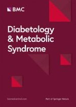
In the absence of Wnt ligands, β-catenin is phosphorylated by a degradation complex composed of Axin, adenomatous polyposis (APC), casein kinase 1 (CK-1), and glycogen synthase kinase 3β (GSK-3β). Subsequently, phosphorylated β-catenin is degraded via the ubiquitin-proteasome pathway. Once a canonical Wnt ligand binds to the dual receptor complex composed of FZD and LRP5/6, the Wnt/β-catenin signaling pathway is activated. Axin subsequently moves to the tail of LRP5/6, while dishevelled (DVL) is recruited by FZD to form a complex with APC, CK-1, and GSK-3β, leading to the accumulation of β-catenin in the cytoplasm [23, 24]. The accumulated β-catenin translocates to the nucleus, whereby it interacts with lymphoid enhancer factor/T cell factor (LEF/TCF) to stimulate the expression of target genes [25, 26]. As agonists of this pathway, Norrie Disease gene product (Norrin) and R-spondin proteins can enhance the canonical Wnt signaling pathway [27]. Studies have confirmed that Wnt/β-catenin signaling is active in various osteoblast and osteoblast precursor cell lines, and can promote bone formation when activated in these cells [28, 29] (Fig. 1).

Effect of high glucose on the canonical wnt signaling pathway in bone homeostasis

Ligands

Wnt1/β-catenin signaling, Wnt3a/β-catenin signaling and Wnt10b/β-catenin signaling axes are critical for the transition of BMSCs [or mouse embryo osteoblast precursor cells (MC3T3-E1 cells)] to the osteoblasts [3035]. Conditional expression of Wnt1 under physiological conditions rapidly increases the number and function of osteoblasts, thus causing a rapid increase in bone mass [36, 37]. Wnt1 inhibits osteoclastogenesis via canonical Wnt signalling in a mouse monocyte-macrophage leukemia cell line (RAW264.7 cells) [38]. Wnt3a, an important target of bone remodeling, increased the secretion of osteoprotegerin [OPG, a decoy receptor of receptor activator of nuclear factor κB ligand (RANKL)] by activating the Wnt/β-catenin signaling pathway to accelerate bone repair processes in osteoporosis models; whereas, silencing of Wnt3a reduced osteoblast differentiation and mineralization [39, 40]. Wnt3a can directly suppresses osteoclast differentiation through canonical Wnt signalling in bone marrow monocyte-macrophage lineage cells (BMMs) [41]. Stevens et al. found that deletion of Wnt10b in mice caused a decrease in the number of mesenchymal progenitor cells rather than altered osteoclast activity or number [42], indicating that Wnt10b functions predominantly on mesenchymal or osteoblast progenitor cells. Increasing expression of Wnt10b was shown to improve glucose homeostasis and reduce plasma triglyceride levels in obese rats [43]. Peroxisome proliferator-activated receptorγ (PPARγ) and CCAAT/enhancer binding proteinα(C/EBPα) are the ultimate regulators in the differentiation of BMSCs to adipocytes, whereas PPARγ and C/EBPα are in a suppressed state when the canonical Wnt signaling pathway is activated [44]. Notably, Wnt1, Wnt3a and Wnt10b are endogenous inhibitors of adipocytogenesis [4547].
However, Shao et al. found that expression of Wnt1, Wnt3a, and Wnt10b in a mouse model of type 2 diabetic osteoporosis established with streptozotocin was significantly lower compared with that in control groups. Moreover, this study found that chronic hyperglycemia and production of advanced glycosylation end products (AGEs) were the main causes of the observed adverse effects [48]. This implies that a decrease in Wnt1, Wnt3a and Wnt10b caused by high glucose can lead to a decrease in osteoblastogenesis, an increase in osteoclastogenesis and adipocytogenesis through canonical Wnt signaling (Figs. 2 and 3).
High dose of Wnt16 can not only promote the expression of OPG by activating the canonical Wnt signaling pathway in osteoblasts, but also accelerate the differentiation process toward osteoblasts by activating the WNT16/β-catenin axis in BMSCs or MC3T3-E1 cells [4951]. In contrast to the findings in osteoblasts, Movérare-Skrtic et al. found that Wnt16 did not significantly regulate β-catenin protein levels or Axin2 expression in mouse osteoclast precursors [52]. Nevertheless, we have not found researches on how Wnt16 affects the differentiation of BMSCs to adipocytes. However, Chen et al. found that Wnt16 expression was significantly reduced in cortical bone of type 1 diabetes mellitus (T1DM) mice. Indeed, this was the main reason for the unexpected increase in cortical porosity and bone resorption in the context of T1DM, and AGEs and oxidative stress were involved in these abnormal changes [53]. This implicates that a decrease in Wnt16 expression can lead to a decrease in osteoblastogenesis through canonical Wnt signaling (Fig. 2).

Receptors

In the physiological state, the two receptors LRP5 and LRP6 are not completely redundant, and both must be expressed in osteoblast lineage for normal bone development. Reducing LRP5/6 protein expression prevents Wnt3a-suppressed differentiation of BMMs to osteoclasts [41]. Augmentation of LRP5/6 expression prevents the differentiation of bone marrow stromal cells of ST2 mice (ST2 cells) into adipocytes [54, 55]. However, Wang et al. found that expression of LRP5 and OPG was significantly reduced in fractures of T1DM rats compared with that in fractures of rats without concomitant diabetes, and bone remodeling could be significantly promoted by inhibiting DKK-1 and improving islet function in this model [56]. Cai et al. found that LRP6 expression was significantly reduced in bone defects of a rabbit model of T1DM compared with that in controls, and confirmed involvement of hyperglycemia-induced inhibition of the canonical Wnt signaling pathway in abnormal bone turnover in T1DM [57].
In addition, promotion of LRP5 expression increased bone mass and bone strength in insulin-deficient diabetic mice, delaying the onset of hyperglycemia in these mice [58]. LRP5 is also essential for normal cholesterol metabolism and glucose-induced insulin secretion. For example, mice specifically lacking LRP5 in osteoblasts and osteocytes exhibited reduced energy expenditure and increased body fat [59]. Male mice with Lrp5 deletion in osteoblasts fed a high-fat diet also showed strong tendencies for hyperglycemia and insulin resistance [60].
FZD is another key receptor involved in canonical Wnt signaling. Kushwaha et al. found that FZD4-deficient mice exhibited a significant decrease in cortical bone mineral density (BMD) and impaired femoral trabecular formation despite a simultaneous increase in FZD8 expression, suggesting that FZD4 remains necessary for normal bone remodeling even in the presence of FZD8 compensation [61]. However, Wang et al. determined that FZD4 expression in BMSCs of diabetic rats was significantly reduced compared with that in controls and triggered the impairment of bone formation. Moreover, the inhibitory effect of hyperglycemia on bone formation was significantly alleviated by targeting expression of FZD4 and β-catenin [62] .
From the above studies, we can confirm that high glucose reduces the expression of LRP5/6 in diabetic individuals to inactivate canonical Wnt signaling, which subsequently inhibits osteoblastogenesis, increases osteoclastogenesis and adipocytogenesis. However, inhibition of osteoblastogenesis due to reduced FZD4 expression caused by high glucose is currently only seen in mesenchymal stem cells (Figs. 2, 3, 4 and 5).

Β-catenin

As a key component of the canonical Wnt signaling pathway, β-catenin is required for postnatal osteoblast differentiation and proliferation, and essential for the anabolic effect of parathyroid hormone in bone [63]. β-catenin deletion at committed stages of osteoclast differentiation will result in osteoporosis because of enhanced osteoclast differentiation [41]. Pharmacological treatments that activate β-catenin in BMSCs can trigger osteoblastogenesis and prevent adipogenesis [64]. However, in an analysis of 40 postmenopausal women with type 2 diabetes mellitus (T2DM) and 40 healthy controls, Gaudio et al. found that β-catenin levels were significantly lower in women with T2DM compared with those in the control group, which may be related to the production of AGEs and increase of SOST [65]. Razny et al. demonstrated that the expression of Wnt pathway antagonists DKK, SOST, SFRPs, and WIF1 was significantly upregulated in obese individuals with T2DM and insulin resistance, while expression of β-catenin (a key factor of osteoblast formation) was significantly decreased [66].
Inhibition of the canonical Wnt signaling pathway mainly targets the degradation of β-catenin. High glucose can also significantly decrease levels of β-catenin in MC3T3-E1 cells by inducing the production of reactive oxygen species (ROS) and AGEs [67]. However, Li et al. found that inhibition of GSK-3β activity in BMSCs of diabetic osteoporotic rats could activate β-catenin and relieve the inhibition of osteoblast differentiation under hyperglycemia [68]. In addition, Xiong et al. demonstrated that osteoblasts treated with Wnt3a could alleviate the inhibition of β-catenin caused by high glucose, and 1,25(OH)2D3 could promote β-catenin transfer from forkhead transcription factor O1 to Wnt/TCF-mediated transcription under high glucose [69].
Collectively, these researches indicate that inhibition of β-catenin activity in diabetic individuals will contribute to reduced bone formation, increased bone resorption, and enhanced adipogenesis. Thus β-catenin is a key factor in reversing the inhibitory state of canonical Wnt signaling (Figs. 2 and 3).

SOST

Specifically secreted by osteocytes, SOST has an antianabolic effect on bone formation and inhibits osteoblast activity to downregulate bone turnover rates [70, 71]. Reduction of the SOST gene can promote bone formation through Wnt/β-catenin signaling and compensate for particle-induced osteolysis [72]. Inhibition of SOST expression resulted in a dramatic reduction in the number of osteoclasts in cancellous bone, thus causing an increase in cancellous bone mass [73]. Sclerostin also attenuated canonical Wnt3a-inhibited adipocyte differentiation [74], showing that SOST is a promotive factor for adipocytogenesis. However, serum levels of SOST are significantly increased in patients with T2DM and femur fractures [75], suggesting that hyperglycemia may increase the expression of SOST by antagonizing canonical Wnt signaling to cause bone resorption beyond bone formation and increase adipocytogenesis. In addition, the osteoclastogenic factor RANKL was positively correlated with SOST expression [76]. Neumann et al. found that serum SOST levels were significantly increased in adults with T1DM compared with those in controls, and a positive correlation between age and serum SOST level was more obvious in these patients [77]. Tsentidis et al. confirmed that SOST was positively correlated with bone formation and resorption markers in children and adolescents with T1DM [78]. Singh et al. found that serum SOST levels were higher in individuals with pre-diabetes and strongly associated with systemic insulin sensitivity, fasting endogenous glucose production, and insulin clearance rate [79]. IDG-SW3 cells (an osteocyte-like cell line) cultured under high glucose conditions displayed significantly increased SOST expression compared with the control group [80].
In conclusion, these results suggest that SOST not only modulates the homeostasis of bone metabolism, but it is crucial for the regulation of human glucose metabolism. Thus, SOST is clearly important for the processes underlying diabetic osteoporosis and deserves greater investigation in DM-induced bone metabolic dysfunction to further elucidate its pathogenic roles (Figs. 2 and 3).

Dkk-1

DKK-1, another antagonist of the Wnt/β-catenin signaling pathway, is predominantly expressed by osteocytes, although not as highly or selectively as SOST. DKK-1 is an important regulator of bone mass, but its overexpression can impair bone regeneration [81]. Heiland et al. found that the number of osteoclasts was significantly reduced in mice treated with anti-DKK-1 antibody [82], suggesting that DKK-1 is a positive regulator of osteoclastogenesis. Knockdown of DKK-1 abrogated dexamethasone-induced accumulation of cytoplasmic oil droplets and the number of adipocyte-like cells in cultured mouse mesenchymal progenitors [83], suggesting that DKK-1 is also a positive regulator of adipocytogenesis. In addition, when DKK1 and RANKL levels were significantly and simultaneously increased, osteoblast differentiation and OPG expression were more inhibited in patients with osteogenesis imperfecta with higher DKK-1 levels [84], demonstrating that DKK-1 has a more detrimental effect on bone formation.
However, in children and adolescents with T1DM, DKK-1 levels were more highly expressed compared with those in controls, thus causing a significant inhibition of the Wnt/β-catenin pathway. The result is reduced osteoblast activation and increased osteoclastogenic factors, which are responsible for reduced bone turnover in individuals with diabetes [85], implicating that hyperglycemia can have a direct negative effect on bone metabolism in young patients by increasing DKK-1 levels. In addition, DKK-1 is positively correlated with fasting plasma glucose, but is mainly seen in children and adolescents with T1DM [85, 86]. In a comparison of 21 women with T2DM and 21 women without diabetes as controls, Sassi et al. found a lower bone turnover rate and significantly higher DKK-1 in women with T2DM [87], indicating that poor glycemic control may interfere with the balance of cytokines associated with normal bone turnover. A significant increase in DKK-1 has also been found in patients with T2DM combined with cardiovascular disease, while a correlation between DKK-1 and vertebral fractures has not been demonstrated in these individuals. In addition, Wnt/β-catenin signaling in a T1DM mouse model induced by streptozotocin was significantly downregulated compared with that in the control group, which was confirmed by increased DKK-1 levels and decreased β-catenin levels in the proximal tibia [88]. Although knockdown of the Dkk-1 gene in osteoblasts did not alter the metabolic parameters of T1DM, it did partially prevent trabecular bone defects and completely prevent cortical bone damage caused by T1DM [89]. Previous studies have also found positive correlations between DKK-1 and SOST in individuals with T2DM [90].
From the findings above, it can be confirmed that DKK-1 is an important negative regulator of bone formation. Increased DKK-1 can trigger increased bone loss and bone marrow adipogenesis in DM and is closely associated with reduced glucose metabolism in children and adolescents with T1DM (Figs. 2 and 3).

SFRPs and WIF1

SFRPs can directly bind to Wnts to block the transmission of Wnt signaling. Notably, the role of SFRP4 in diabetic osteoporosis is perhaps the most unique. Impaired osteoblast differentiation was accompanied by upregulation of the Wnt signaling antagonist SFRP4 and impairment of Wnt signaling [91]. The loss of canonical Wnt signaling pathway inhibition in SFRP4-KO mice caused increases in bone trabecular bone mass [92]. Enhanced adipogenic differentiation can be inhibited in ST2 cells by silencing SFRP4 to activate canonical Wnt signaling [93], indicating that SFRP4 is a promoter of adipocytogenesis. The increase of bone fragility induced by oxidative stress caused by diabetes and aging is also partly related to the inhibition of Wnt signaling through upregulation of SFRP4 [94]. In addition, Diabetic osteoporosis caused by decreased Wnt/β-catenin signaling is partly induced by epigenetic derepression of the SFRP-4 gene [95]. Mastaitis et al. found that diet-induced obese SFRP4-KO mice exhibited reduced energy expenditure, food intake, and BMD, but had similar glucose tolerances to control mice; moreover, SFRP4 overexpression did not affect energy metabolism or glucose homeostasis [96]. Because SFRP-4 is a soluble factor, the use of neutralizing antibodies or inhibitors could be a good therapeutic strategy for diabetic osteoporosis [95]. Altogether, these results indicate that hyperglycemia-induced elevation of SFRP4 leads to an imbalance in bone remodeling and increased adipocytogenesis, rather than glucose metabolism. Therefore, SFRP4 may be a good target for the treatment of diabetic osteoporosis (Figs. 2 and 3).
Like SFRPs, WIF1 also acts as a decoy receptor and inhibits canonical Wnt signaling. The WIF1/Wnt/β-catenin pathway plays an essential role in osteogenic differentiation [97]. Morimoto et al. found that glucocorticoid-mediated inhibition of osteoblast growth and reduction of differentiation of ST2 cells to osteoblasts could be alleviated by silencing WIF1 [98], demonstrating that WIF1 is an inhibitory factor of osteoblastogenesis. WIF1 is elevated in individuals with T2DM [66]. Nevertheless, currently there is a lack of WIF1 effects on osteoclastogenesis and adipocytogenesis through canonical Wnt signaling. However, it is at least certain that elevation of WIF1 in T2DM individuals inhibits osteoblast differentiation and growth via canonical Wnt signaling (Fig. 2).
Altogether, the results described above demonstrate that in addition to affecting bone homeostasis, the Wnt/β-catenin signaling pathway is closely related to endocrine metabolism. Moreover, the Wnt/β-catenin signaling pathway is significantly inhibited in the high glucose state, which leads to reduced bone formation, enhanced bone resorption, elevated bone marrow adipogenesis, inhibited insulin secretion, and reduced glucose and lipid metabolism.

Non-canonical wnt signaling pathway

The non-canonical Wnt signaling pathway is involved in morphological maturation caused by osteoblast and osteoclast differentiation because it plays important roles in the regulation of cell motility, cell polarity, and cytoskeleton remodeling [99]. The non-canonical Wnt signaling pathway can be divided into the Wnt/planar cell polarity (PCP) signaling pathway and Wnt/Ca2+ signaling pathway. In the Wnt/PCP signaling pathway, a complex formed by FZD and RYK or ROR1/2 binds to Wnt proteins and co-recruits DVL, which then binds to dishevelled associated activator of morphogenesis 1 (DAAM1) to co-activate Rac family small GTPase1 (RAC1), RAS homolog family member A (RhoA), and cell division cyclin 42 (CDC42). Activation of these Rho family small GTPases is involved in the regulation of cytoskeleton remodeling, actin polymerization, cell polarization, and cell motility. Among them, Rac1 and CDC42 can co-activate c-Jun N-terminal kinase (JNK) to participate in cell migration and cell polarization. JNK subsequently activates transcription factors such as Jun proto-oncogene (c-Jun) and activator protein 1 (AP-1) to regulate downstream gene expression. RhoA is involved in activation of RHO-associated kinase (ROCK), which regulates actin polymerization and cytoskeleton remodeling (Fig. 1).
In the Wnt/Ca2+ signaling pathway, the complex formed by FZD and RYK or ROR1/2 can also activate phospholipase C (PLC) by activating G proteins on the cell membrane after binding to Wnt proteins. Activated PLC can decompose phosphatidylinositol-4,5-bisphosphate (PIP2) into 1,2-diacylglycerol (DAG) and inositol-1,4,5-trisphosphate (IP3) on the cell membrane. IP3 induces the release of large amounts of Ca2+ into the cytoplasm through IP3-sensitive Ca2+ channels in the endoplasmic reticulum. At this point, the decrease of intracellular Ca2+ store leads to the opening of Ca2+ channels regulated by Ca2+ reservoirs on the cell membrane, which causes a large influx of Ca2+ into the cytoplasm, resulting in a transient increase in free cytoplasmic Ca2+. In concert with Ca2+, DAG can also activate protein kinase C (PKC), which activates CDC42 to participate in morphological maturation processes such as cytoskeleton remodeling. Ca2+ can simultaneously activate calcineurin, which once activated can dephosphorylate nuclear factor-activated T cell (NFATc) protein. Subsequently, the dephosphorylated NFATc protein is transferred to the nucleus to form NFAT complexes (NFATn) with specific transcription factors to regulate the expression of downstream target genes. In addition, Ca2+ activates calmodulin-dependent protein kinase II (CaMKII), which phosphorylates transforming growth factor (TGF)-β-activated kinase 1 (TAK-1) to induce activation of the transcription factor Nemo-like kinase (NLK). NLK can act as an inhibitor of the canonical Wnt signaling pathway by reducing binding of β-catenin to TCF/LEF [100] (Fig. 1).

Effect of high glucose on non-canonical wnt signaling in bone homeostasis

Ligands

Although Wnt3a is usually considered a ligand for activation of the canonical Wnt signaling pathway, studies have shown that it can also mediate osteoblastogenesis independently of β-catenin by activating the heterotrimeric G protein Gα(q/11)/PLC/PKC pathway in ST2 cells or JNK pathway in human mesenchymal stem cells (hMSCs) [101, 102]. However, Wnt3a expression was significantly decreased in a mouse model of type 2 diabetic osteoporosis [48], suggesting that the reduction of Wnt3a in a high-glucose state could inhibit bone formation by affecting the non-canonical Wnt signaling pathway in these cells. Insulin can promote bone formation. As a ligand of the non-canonical Wnt signaling pathway, Wnt4 can completely antagonize Wnt3a-stimulated cell growth and insulin secretion in a rat islet cell tumor cell line (INS-1) [103]. However, Wnt4 expression in islet cells of individuals with diabetes is increased, leading to enhancement of the JNK pathway in Wnt/PCP signaling, which impairs β cell proliferation and insulin secretion [104]. Thus, the increase of Wnt4 in individuals with diabetes can indirectly affect the osteogenesis process by affecting insulin synthesis (Fig. 4).
Wnt5a can increase osteoblast differentiation by activating the Ror2/ROCK pathway in hMSCs [105]. However, Xu et al. observed a trend for increased Wnt5a levels in individuals with long-term T2DM or T2DM after 3 months of treatment. Accordingly, the authors suggest that this may be one of the reasons for the elevated BMD in individuals with T2DM [106]. However, bone strength and turnover are low in individuals with T2DM [107] due to osteoblast-expressed Wnt5a accelerating the differentiation of monocytes into osteoclasts [108]. Furthermore, in osteoclast precursors, Wnt5a also promotes the binding of RANKL to receptor activator of nuclear factor-κB (RANK) by binding to ROR2 [109]. Although the binding of RANKL to RANK will sequentially activate tumor necrosis factor-associated factor 6 (TRAF6) and nuclear factor (NF)-κB, the latter can regulate osteoclast formation by activating downstream NFATc1. However, Zhang et al. found that the canonical Wnt signaling pathway was significantly inhibited in a rat model of type 2 diabetic osteoporosis, while the RANKL-activated TRAF6/NF-κB/NFATc1 pathway was significantly activated, resulting in increased osteoclast differentiation [110]. These results suggest that high glucose can also promote bone resorption through this pathway. Razny et al. observed increased expression of RANK and RANKL in obese individuals with T2DM and insulin resistance [66]. RANKL levels were also significantly elevated in children and adolescents with T1DM [111]. Ding et al. confirmed that high glucose could enhance the differentiation of RAW264.7 cells into osteoclasts by increasing the RANKL/OPG ratio. Moreover, the enhancement of bone resorption was caused by high glucose inducing the production and accumulation of AGEs in vivo [112]. After applying a combination of high glucose plus palmitate to mimic the effects of the microenvironment of T2DM on osteoclast differentiation, Qu et al. found that this environment promoted differentiation of osteoclast precursors into osteoclasts by decreasing OPG expression and increasing levels of RANK, RANKL, and NFATc1. Moreover, this study found that attenuating the activity of p66Shc both inhibited ROS production and reversed the increase in osteoclastogenesis caused by high glucose and palmitate [113]. In summary, although elevating Wnt5a in a hyperglycemic state can increase BMD, this alone is insufficient to fully assess the risk of fracture in individuals with diabetes [114]. In addition, elevated Wnt5a enhances the binding of RANK to RANKL, leading to increased osteoclast production. Hyperglycemia can decrease expression of OPG, the main decoy receptor of RANKL, leading to an increase in RANKL. Therefore, the authors concluded that the elevation of Wnt5a caused by long-term hyperglycemia will eventually elicit a negative balance of bone homeostasis (Fig. 5).
Keats et al. demonstrated that high glucose significantly increased expression of Wnt11, another ligand of the non-canonical Wnt signaling pathway, in human bone marrow mesenchymal progenitor cells (MPCs), which promoted their differentiation towards adipocytes rather than osteoblasts through the Wnt11/PKC pathway [115]. Wnt16 promoted osteoblast formation through the transcription factors recombinant FOS-like antigens 1 and 2 (Fosl1 and Fosl2), which are activated by the non-canonical Wnt/JNK pathway in primary osteoblasts [116]. Wnt16 not only inhibits osteoclastogenesis by blocking the binding of RANKL to RANK, it reduces osteoclast differentiation by inhibiting RANKL-induced activation of NF-κB and NFATC1 by activating the non-canonical Wnt signaling pathway in osteoclast precursors. In addition, Maeda et al. observed normal bone mass of cancellous bone and significantly reduced bone mass of cortical bone in Wnt16-knockout mice, indicating that Wnt16 expression in cortical bone was higher than in cancellous bone [117]. However, compared with that in the control group, Wnt16 expression was significantly reduced in a type 1 diabetic osteoporosis mouse model, leading to a significant increase in the cortical bone porosity and bone resorption. Mechanistically, the accumulation of oxidative stress caused by hyperglycemia was implicated in these observed abnormalities [53] (Figs. 4 and 5). Notably, the findings of this study also explain the increased presence of cortical bone defects [89] in adults with T1DM.

Receptors

Both ROR1 and ROR2 are essential for bone development. However, the prevailing view is that only ROR2 affects bone mass. Maeda et al. demonstrated that Wnt5a-ror2 signaling in osteoclast precursors can enhance RANKL-induced osteoclastogenesis by activating JNK and recruiting c-Jun to the RANK gene promoter [117]. However, there is a lack of studies on the effect of ROS on ROR2 expression in osteoclast precursors. Lee et al. showed that ROS can cause periodontitis by upregulating expression of Wnt5a, FZD, and ROR2 in human periodontal ligament cells [118]. As mentioned above, Wnt5a expression is increased in individuals with diabetes. Therefore, ROS produced in response to high glucose appears to activate Wnt5a-ROR2 signaling in osteoclast precursors to increase osteoclast differentiation.

SFRPs and WIF1

Although SFRP4 can reduce osteoclast formation and reduce osteoclast activity by inhibiting the non-canonical Wnt/Ror2/JNK cascade [119], Haraguchi et al. demonstrated that persistent elevation and activation of SFRP4 could unlock the inhibitory effect of canonical Wnt signaling on osteoclastogenesis, resulting in low bone mass [94]. In addition, SFRP4 has been be closely associated with insulin resistance and the progression of T2DM. SFRP4 expression is significantly higher in patients with prediabetes, T2DM, or gestational diabetes compared with that in healthy individuals [120122]. Mori et al. also found that treatment of ST2 cells with methylglyoxal (a glucose intermediate metabolite) rapidly enhanced SFRP4 expression and inhibited expression of osteogenic Wnt-targeting genes, including OPG [95]. Taken together, the elevation of SFRP4 caused by high glucose ultimately has a detrimental effect on bone formation (Fig. 5).
WIFI also inhibits non-canonical Wnt signaling pathway. WIF1 is elevated in individuals with T2DM [66], however, there is a lack of researches related to WIF1 affecting osteoblastogenesis, osteoclastogenesis, or adipocytogenesis through non-canonical Wnt signaling. Therefore, we believe that exploring high glucose to affect bone remodeling via non-canonical Wnt signaling, WIF1 will have the potential to be one of the key factors in the future.

Wnt/planar cell polarity signaling pathway

Although differentiation of osteoblasts requires involvement of the RhoA/ROCK pathway [123], its activation can have a significant negative regulatory effect on osteoblastogenesis [124]. Moreover, activation of the RhoA/ROCK pathway inhibits osteoclast formation by osteoclast precursors [125]. However, there is a lack of data describing the effects of the RhoA/ROCK pathway on osteoblastogenesis and osteoclastogenesis in a high-glucose state. Cheng et al. showed that high glucose induces activation of the RhoA/ROCK pathway in RAW264.7 cells [126]. Activation of the RhoA/ROCK pathway is an important cause of long-term complications of diabetes [127]. Therefore, high glucose may activate the RhoA/ROCK pathway in both osteoblast and osteoclast precursors, thereby inhibiting bone formation and resorption; however, more studies are needed. It is certain that restoring the normal functioning of RhoA/ROCK by controlling hyperglycemia can be an important target for the treatment of diabetic complications, including diabetic osteoporosis.
Rac1 and Cdc42 are involved in bone resorption processes such as osteoclast polarization and inhibition of osteoclast apoptosis. Umbayev et al. [128] demonstrated that inhibition of Cdc42 levels increased differentiation of rat adipose-derived mesenchymal stem cells into osteoblasts and decreased ROS levels in these cells. When RANKL-induced osteoclast differentiation and bone resorption are inhibited, the activities of Rac1 and JNK are simultaneously diminished [129], suggesting a positive feedback effect of the Rac1/JNK cascade in osteoclastogenesis and role in bone resorption. In general, ROS is considered a pathological factor in inflammation and aging. For example, Lee et al. found that RANKL-stimulated BMMs transiently increased intracellular ROS levels by activating signaling cascades, including Rac1 and JNK, and demonstrated that ROS can act as a physiological second messenger in RANKL signaling to promote osteoclast formation [130]. Specifically, Rac1 was closely associated with ROS production [131]. Thus, ROS produced by high glucose can ultimately increase osteoclast differentiation by activating the Rac1/JNK cascade in osteoclast precursors (Fig. 5). Rac1 is also important for the nuclear translocation of β-catenin during osteoblastogenesis. Although there is a lack of data regarding the effect of high glucose on Rac1 in osteoblast precursors or immature osteoblasts, it has been shown that silencing Rac1 limits ROS production to protect cells from ROS-induced damage and apoptosis [132]. Accordingly, Rac1 may also be an important therapeutic target for reducing osteoblast damage caused by high glucose-induced oxidative stress. Activation of the JNK pathway has a facilitative effect on differentiation of hMSCs into osteoblasts and inhibits adipocyte production [133]. However, Wnt signaling factors associated with the non-canonical pathway (i.e., Wnt5a/b, Ror1, Ror2, whole and phosphorylated JNK, whole and phosphorylated c-Jun) were almost constant in ST2 cells treated with methylglyoxal compared with those in control ST2 cells [95]. Furthermore, Chaves et al. demonstrated that high glucose failed to increase JNK phosphorylation in BMSCs from young Wistar and spontaneous hypertensive rats without hypertension. However, the concentration of glucose used in the experiment was insufficient to cause more severe damage to BMSCs, which the authors note may have contributed to the results of the study [134]. Thus, the effect of high glucose on the Rac1/JNK pathway in osteoblast precursors and immature osteoblasts needs further investigation.

Wnt/Ca2+ signaling pathway

Strack et al. found that the Gα(q/11)/PLC/PKC pathway is similar to the RANKL/RANK pathway, and both can promote the differentiation of osteoclast precursors into osteoclasts [135]. The Gα(q/11)/PLC/PKC pathway can promote differentiation of ST2 cells into osteoblasts. ROS produced by high glucose has been found to activate PKC by increasing free Ca2+ concentrations in the cytoplasm, thereby enhancing bone morphogenic protein 2-induced collagen synthesis and early osteogenic gene expression in rat spinal ligament cells [136]. However, because there is a current lack of studies on the effects of high glucose on bone formation or bone resorption through this pathway, it represents a new research direction for diabetic osteoporosis.
The Ca2+/CaMKII/TAK-1/NLK pathway inhibits the transactivation of peroxisome proliferator-activated receptor γ and induces Runx2 expression, thereby promoting the differentiation of bone marrow mesenchymal progenitors towards osteoblasts rather than adipocytes. However, Li et al. found that NLK expression was significantly increased in BMSCs from type 2 diabetic Goto-Kakizaki rats, which in turn triggered TCF inactivation to inhibit the bone formation process induced by the canonical Wnt signaling pathway. Again, the reported abnormalities were closely associated with the production of AGEs and insulin resistance [137] (Fig. 4). Ca2+ concentrations and CaMKII phosphorylation were significantly increased in osteoclasts differentiated from RAW264.7 cells treated with macrophage colony-stimulating factor and RANKL [138], implicating the Ca2+/CaMKII pathway in osteoclast differentiation. However, Shen et al. found that hyperglycemia-induced oxidative stress can increase osteoclastogenesis and bone resorption in type 1 diabetic rats by increasing free Ca2+ concentrations in BMMs, thus activating the Ca2+/CaMKII pathway. Moreover, application of KN93 (a CaMKII inhibitor) significantly alleviated the hyperglycemia-induced abnormal bone morphology in rats but failed to reduce hyperglycemia and body weight [139]. Activation of CaMKII can stimulate the TAK-1/NLK pathway. Thus, based on the information above, it can be concluded that high glucose can induce activation of the Ca2+/CaMKII/TAK-1/NLK pathway in osteoclast precursors and increase SFRP4 expression. The result is more severe inhibition of the Wnt/β-catenin pathway in these cells, which increases osteoclast formation to further aggravate bone resorption (Fig. 5).
The Ca2+/Calcineurin/NFATc1 pathway plays a key role in the negative regulation of osteoblast differentiation. Vadavanath et al. showed that ROS can activate the Ca2+/calcineurin/NFATc1 pathway to regulate the expression of specific transcription factors [140]. However, Jie et al. found that application of FK506 (a calcineurin inhibitor) significantly alleviated the detrimental effects of oxidative stress on bone formation by activating the Ca2+/Calcineurin/NFATc1 pathway in MC3T3-E1 cells [141]. These results indicate that in a high-glucose state, ROS production activates the Ca2+/Calcineurin/NFATc1 pathway in osteoblast precursors to inhibit their differentiation into osteoblasts (Fig. 4). Moreover, the Ca2+/calcineurin/NFATc1 pathway is particularly critical in the process of osteoclast differentiation [142]. In this pathway, NFATc1 acts a key factor for osteoclast differentiation; indeed, even in the absence of osteoclast differentiation factors, ectopic overexpression of NFATc1 is sufficient to induce osteoclast differentiation [143]. However, Kim et al. found that ROS can promote the differentiation of bone marrow-derived monocytes into osteoclasts by enhancing the PLCγ/Ca2+/NFATc1 pathway [144]. Collectively, these results suggest that increased ROS production in response to high glucose can accelerate osteoclast differentiation by activating the PLCγ/Ca2+/NFATc1 pathway in osteoclast precursors (Fig. 5).
Based on the findings described above, we conclude that abnormalities of the non-canonical Wnt pathway will affect the morphological maturation of many cell types. Hyperglycemia not only disturbs this pathway by affecting the expression of ligands and receptors, it can directly interfere with the PCP or Ca2+ pathways of these cells by producing ROS and AGEs. Thus, if hyperglycemia is prolonged, the effects on affected cells are more harmful and inhibiting CaMKII or calcineurin activity may be a new therapeutic target for diabetic osteoporosis.

Conclusion

Therefore, it can be concluded that high glucose affects bone homeostasis by disrupting the Wnt pathway. In addition, high glucose can interfere with the normal bone remodeling process by affecting multiple cell types simultaneously. In this process, production of ROS and AGEs, as well as the development of insulin resistance, are critical disruptors of the Wnt pathway. Hyperglycemia can inhibit bone formation and promote bone resorption by suppressing the canonical Wnt signaling pathway. In addition, the non-canonical Wnt signaling pathway can indirectly lead to impaired bone formation by inhibiting insulin secretion and promoting the differentiation of MPCs into adipocytes. Compared with the canonical Wnt pathway, alterations in levels of other Wnt ligands, the degree of Ror2 receptor expression, and interference with the Wnt/PCP pathway by hyperglycemia have not been studied enough. Therefore, more attention should be paid in the future to the effects of hyperglycemia on bone homeostasis through the non-canonical Wnt pathway, and more clinical trials should be conducted accordingly. This strategy will bring about more possibilities for designing new drugs to treat diabetic osteoporosis by interfering with the non-canonical Wnt signaling pathway.

Acknowledgements

We thank Liwen Bianji(Edanz) (www.​liwenbianji.​cn/​) for basic language editing of a draft of this manuscript.

Declarations

Conflict of interest

Kairan Bao, Yinghua Jiao, Lei Xing, Fang Zhang and Faming Tian declared no conficts of interest.

Ethical approval

Not applicable.
Open AccessThis article is licensed under a Creative Commons Attribution 4.0 International License, which permits use, sharing, adaptation, distribution and reproduction in any medium or format, as long as you give appropriate credit to the original author(s) and the source, provide a link to the Creative Commons licence, and indicate if changes were made. The images or other third party material in this article are included in the article’s Creative Commons licence, unless indicated otherwise in a credit line to the material. If material is not included in the article’s Creative Commons licence and your intended use is not permitted by statutory regulation or exceeds the permitted use, you will need to obtain permission directly from the copyright holder. To view a copy of this licence, visit http://​creativecommons.​org/​licenses/​by/​4.​0/​. The Creative Commons Public Domain Dedication waiver (http://​creativecommons.​org/​publicdomain/​zero/​1.​0/​) applies to the data made available in this article, unless otherwise stated in a credit line to the data.

Publisher’s Note

Springer Nature remains neutral with regard to jurisdictional claims in published maps and institutional affiliations.
Literatur
1.
Zurück zum Zitat American Diabetes Association Professional Practice Committee. 2. Classification and diagnosis of diabetes: Standards of Medical Care in Diabetes-2022. Diabetes Care. 2022;45(Suppl 1):17–S38.CrossRef American Diabetes Association Professional Practice Committee. 2. Classification and diagnosis of diabetes: Standards of Medical Care in Diabetes-2022. Diabetes Care. 2022;45(Suppl 1):17–S38.CrossRef
2.
Zurück zum Zitat Vokó Z, Gáspár K, Inotai A, Horváth C, Bors K, Speer G, et al. Osteoporotic fractures may impair life as much as the complications of diabetes. J Eval Clin Pract. 2017;23(6):1375–80.PubMedCrossRef Vokó Z, Gáspár K, Inotai A, Horváth C, Bors K, Speer G, et al. Osteoporotic fractures may impair life as much as the complications of diabetes. J Eval Clin Pract. 2017;23(6):1375–80.PubMedCrossRef
3.
Zurück zum Zitat Chandran M, Tay D, Huang XF, Hao Y. The burden of inpatient care for diabetic and non-diabetic patients with osteoporotic hip fractures-does it differ? An analysis of patients recruited into a fracture liaison service in Southeast Asia. Arch Osteoporos. 2018;13(1):27.PubMedCrossRef Chandran M, Tay D, Huang XF, Hao Y. The burden of inpatient care for diabetic and non-diabetic patients with osteoporotic hip fractures-does it differ? An analysis of patients recruited into a fracture liaison service in Southeast Asia. Arch Osteoporos. 2018;13(1):27.PubMedCrossRef
4.
5.
Zurück zum Zitat Gassel LC, Schneider S, Banke IJ, Braun KF, Volkering C, Zeeb L, et al. Dysregulation of wnt signaling in bone of type 2 diabetes mellitus and diabetic Charcot arthropathy. BMC Musculoskelet Disord. 2022;23(1):365.PubMedPubMedCentralCrossRef Gassel LC, Schneider S, Banke IJ, Braun KF, Volkering C, Zeeb L, et al. Dysregulation of wnt signaling in bone of type 2 diabetes mellitus and diabetic Charcot arthropathy. BMC Musculoskelet Disord. 2022;23(1):365.PubMedPubMedCentralCrossRef
6.
Zurück zum Zitat Cosin-Roger J, Ortiz-Masià MD, Barrachina MD. Macrophages as an emerging source of wnt ligands: relevance in Mucosal Integrity. Front Immunol. 2019;10:2297.PubMedPubMedCentralCrossRef Cosin-Roger J, Ortiz-Masià MD, Barrachina MD. Macrophages as an emerging source of wnt ligands: relevance in Mucosal Integrity. Front Immunol. 2019;10:2297.PubMedPubMedCentralCrossRef
7.
Zurück zum Zitat Li Z, Yuan X, Arioka M, Bahat D, Sun Q, Chen J, et al. Pro-osteogenic Effects of WNT in a mouse model of bone formation around femoral implants. Calcif Tissue Int. 2021;108(2):240–51.PubMedCrossRef Li Z, Yuan X, Arioka M, Bahat D, Sun Q, Chen J, et al. Pro-osteogenic Effects of WNT in a mouse model of bone formation around femoral implants. Calcif Tissue Int. 2021;108(2):240–51.PubMedCrossRef
8.
Zurück zum Zitat Appelman-Dijkstra NM, Papapoulos SE. Clinical advantages and disadvantages of anabolic bone therapies targeting the WNT pathway. Nat Rev Endocrinol. 2018;14(10):605–23.PubMedCrossRef Appelman-Dijkstra NM, Papapoulos SE. Clinical advantages and disadvantages of anabolic bone therapies targeting the WNT pathway. Nat Rev Endocrinol. 2018;14(10):605–23.PubMedCrossRef
9.
Zurück zum Zitat Janssen R, Pechmann M, Turetzek N. A chelicerate wnt gene expression atlas: novel insights into the complexity of arthropod wnt-patterning. Evodevo. 2021;9(1):12.CrossRef Janssen R, Pechmann M, Turetzek N. A chelicerate wnt gene expression atlas: novel insights into the complexity of arthropod wnt-patterning. Evodevo. 2021;9(1):12.CrossRef
10.
Zurück zum Zitat Yin P, Wang W, Zhang Z, Bai Y, Gao J, Zhao C. Wnt signaling in human and mouse breast cancer: focusing on wnt ligands, receptors and antagonists. Cancer Sci. 2018;109(11):3368–75.PubMedPubMedCentralCrossRef Yin P, Wang W, Zhang Z, Bai Y, Gao J, Zhao C. Wnt signaling in human and mouse breast cancer: focusing on wnt ligands, receptors and antagonists. Cancer Sci. 2018;109(11):3368–75.PubMedPubMedCentralCrossRef
11.
Zurück zum Zitat Kessenbrock K, Smith P, Steenbeek SC, Pervolarakis N, Kumar R, Minami Y, et al. Diverse regulation of mammary epithelial growth and branching morphogenesis through noncanonical wnt signaling. Proc Natl Acad Sci U S A. 2017;114(12):3121–6.PubMedPubMedCentralCrossRef Kessenbrock K, Smith P, Steenbeek SC, Pervolarakis N, Kumar R, Minami Y, et al. Diverse regulation of mammary epithelial growth and branching morphogenesis through noncanonical wnt signaling. Proc Natl Acad Sci U S A. 2017;114(12):3121–6.PubMedPubMedCentralCrossRef
12.
Zurück zum Zitat Nusse R, Clevers H. Wnt/β-Catenin signaling, Disease, and emerging therapeutic modalities. Cell. 2017;169(6):985–99.PubMedCrossRef Nusse R, Clevers H. Wnt/β-Catenin signaling, Disease, and emerging therapeutic modalities. Cell. 2017;169(6):985–99.PubMedCrossRef
13.
Zurück zum Zitat Nie X, Wei X, Ma H, Fan L, Chen WD. The complex role of wnt ligands in type 2 diabetes mellitus and related complications. J Cell Mol Med. 2021;25(14):6479–95.PubMedPubMedCentralCrossRef Nie X, Wei X, Ma H, Fan L, Chen WD. The complex role of wnt ligands in type 2 diabetes mellitus and related complications. J Cell Mol Med. 2021;25(14):6479–95.PubMedPubMedCentralCrossRef
15.
Zurück zum Zitat Ackers I, Malgor R. Interrelationship of canonical and non-canonical wnt signalling pathways in chronic metabolic diseases. Diab Vasc Dis Res. 2018;15(1):3–13.PubMedCrossRef Ackers I, Malgor R. Interrelationship of canonical and non-canonical wnt signalling pathways in chronic metabolic diseases. Diab Vasc Dis Res. 2018;15(1):3–13.PubMedCrossRef
16.
Zurück zum Zitat Chen H, Huang H, Wang Y, Zhang Y, Liu M, Lou Y, et al. Decreased serum wnt antagonist levels in patients with active acromegaly. Endocr Pract. 2022;28(5):515–20.PubMedCrossRef Chen H, Huang H, Wang Y, Zhang Y, Liu M, Lou Y, et al. Decreased serum wnt antagonist levels in patients with active acromegaly. Endocr Pract. 2022;28(5):515–20.PubMedCrossRef
17.
18.
Zurück zum Zitat Baksh D, Tuan RS. Canonical and non-canonical wnts differentially affect the development potential of primary isolate of human bone marrow mesenchymal stem cells. J Cell Physiol. 2007;212(3):817–26.PubMedCrossRef Baksh D, Tuan RS. Canonical and non-canonical wnts differentially affect the development potential of primary isolate of human bone marrow mesenchymal stem cells. J Cell Physiol. 2007;212(3):817–26.PubMedCrossRef
19.
Zurück zum Zitat Alharbi MA, Zhang C, Lu C, Milovanova TN, Yi L, Ryu JD, et al. FOXO1 deletion reverses the Effect of Diabetic-Induced impaired Fracture Healing. Diabetes. 2018;67(12):2682–94.PubMedPubMedCentralCrossRef Alharbi MA, Zhang C, Lu C, Milovanova TN, Yi L, Ryu JD, et al. FOXO1 deletion reverses the Effect of Diabetic-Induced impaired Fracture Healing. Diabetes. 2018;67(12):2682–94.PubMedPubMedCentralCrossRef
20.
Zurück zum Zitat Yang J, Chen S, Zong Z, Yang L, Liu D, Bao Q, et al. The increase in bone resorption in early-stage type I diabetic mice is induced by RANKL secreted by increased bone marrow adipocytes. Biochem Biophys Res Commun. 2020;525(2):433–9.PubMedCrossRef Yang J, Chen S, Zong Z, Yang L, Liu D, Bao Q, et al. The increase in bone resorption in early-stage type I diabetic mice is induced by RANKL secreted by increased bone marrow adipocytes. Biochem Biophys Res Commun. 2020;525(2):433–9.PubMedCrossRef
21.
Zurück zum Zitat Morinaga H, Muta Y, Tanaka T, Tanabe M, Hamaguchi Y, Yanase T. High-mobility group box 2 protein is essential for the early phase of adipogenesis. Biochem Biophys Res Commun. 2021;557:97–103.PubMedCrossRef Morinaga H, Muta Y, Tanaka T, Tanabe M, Hamaguchi Y, Yanase T. High-mobility group box 2 protein is essential for the early phase of adipogenesis. Biochem Biophys Res Commun. 2021;557:97–103.PubMedCrossRef
22.
Zurück zum Zitat Arnold KM, Pohlig RT, Sims-Mourtada J. Co-activation of hedgehog and wnt signaling pathways is associated with poor outcomes in triple negative breast cancer. Oncol Lett. 2017;14(5):5285–92.PubMedPubMedCentral Arnold KM, Pohlig RT, Sims-Mourtada J. Co-activation of hedgehog and wnt signaling pathways is associated with poor outcomes in triple negative breast cancer. Oncol Lett. 2017;14(5):5285–92.PubMedPubMedCentral
23.
Zurück zum Zitat He J, Zhang N, Zhang J, Jiang B, Wu F. Migration critically meditates osteoblastic differentiation of bone mesenchymal stem cells through activating canonical wnt signal pathway. Colloids Surf B Biointerfaces. 2018;171:205–13.PubMedCrossRef He J, Zhang N, Zhang J, Jiang B, Wu F. Migration critically meditates osteoblastic differentiation of bone mesenchymal stem cells through activating canonical wnt signal pathway. Colloids Surf B Biointerfaces. 2018;171:205–13.PubMedCrossRef
24.
Zurück zum Zitat Nelson AL, Fontana G, Miclau E, Rongstad M, Murphy W, Huard J, et al. Therapeutic approaches to activate the canonical wnt pathway for bone regeneration. J Tissue Eng Regen Med. 2022;16(11):961–76.PubMedPubMedCentralCrossRef Nelson AL, Fontana G, Miclau E, Rongstad M, Murphy W, Huard J, et al. Therapeutic approaches to activate the canonical wnt pathway for bone regeneration. J Tissue Eng Regen Med. 2022;16(11):961–76.PubMedPubMedCentralCrossRef
25.
Zurück zum Zitat Wang B, Rong X, Zhou Y, Liu Y, Sun J, Zhao B, et al. Eukaryotic initiation factor 4A3 inhibits Wnt/β-catenin signaling and regulates axis formation in zebrafish embryos. Development. 2021;148(9):dev198101.PubMedCrossRef Wang B, Rong X, Zhou Y, Liu Y, Sun J, Zhao B, et al. Eukaryotic initiation factor 4A3 inhibits Wnt/β-catenin signaling and regulates axis formation in zebrafish embryos. Development. 2021;148(9):dev198101.PubMedCrossRef
26.
Zurück zum Zitat Puzik K, Tonnier V, Opper I, Eckert A, Zhou L, Kratzer MC, et al. Lef1 regulates caveolin expression and caveolin dependent endocytosis, a process necessary for Wnt5a/Ror2 signaling during Xenopus gastrulation. Sci Rep. 2019;9(1):15645.PubMedPubMedCentralCrossRef Puzik K, Tonnier V, Opper I, Eckert A, Zhou L, Kratzer MC, et al. Lef1 regulates caveolin expression and caveolin dependent endocytosis, a process necessary for Wnt5a/Ror2 signaling during Xenopus gastrulation. Sci Rep. 2019;9(1):15645.PubMedPubMedCentralCrossRef
28.
Zurück zum Zitat Du JH, Lin SX, Wu XL, Yang SM, Cao LY, Zheng A, et al. The function of wnt ligands on Osteocyte and Bone Remodeling. J Dent Res. 2019;98(8):930–8.PubMedCrossRef Du JH, Lin SX, Wu XL, Yang SM, Cao LY, Zheng A, et al. The function of wnt ligands on Osteocyte and Bone Remodeling. J Dent Res. 2019;98(8):930–8.PubMedCrossRef
29.
Zurück zum Zitat Lawson LY, Brodt MD, Migotsky N, Chermside-Scabbo CJ, Palaniappan R, Silva MJ. Osteoblast-specific wnt secretion is required for skeletal Homeostasis and Loading-Induced bone formation in adult mice. J Bone Miner Res. 2022;37(1):108–20.PubMedCrossRef Lawson LY, Brodt MD, Migotsky N, Chermside-Scabbo CJ, Palaniappan R, Silva MJ. Osteoblast-specific wnt secretion is required for skeletal Homeostasis and Loading-Induced bone formation in adult mice. J Bone Miner Res. 2022;37(1):108–20.PubMedCrossRef
30.
Zurück zum Zitat Zhang B, Li R, Wang W, Zhou X, Luo B, Zhu Z, et al. Effects of WNT1 c.110 T > C and c.505G > T mutations on osteoblast differentiation via the WNT1/β-catenin signaling pathway. J Orthop Surg Res. 2021;16(1):359.PubMedPubMedCentralCrossRef Zhang B, Li R, Wang W, Zhou X, Luo B, Zhu Z, et al. Effects of WNT1 c.110 T > C and c.505G > T mutations on osteoblast differentiation via the WNT1/β-catenin signaling pathway. J Orthop Surg Res. 2021;16(1):359.PubMedPubMedCentralCrossRef
31.
Zurück zum Zitat Chen Y, Chen L, Yin Q, Gao H, Dong P, Zhang X, et al. Reciprocal interferences of TNF-α and Wnt1/β-catenin signaling axes shift bone marrow-derived stem cells towards osteoblast lineage after ethanol exposure. Cell Physiol Biochem. 2013;32(3):755–65.PubMedCrossRef Chen Y, Chen L, Yin Q, Gao H, Dong P, Zhang X, et al. Reciprocal interferences of TNF-α and Wnt1/β-catenin signaling axes shift bone marrow-derived stem cells towards osteoblast lineage after ethanol exposure. Cell Physiol Biochem. 2013;32(3):755–65.PubMedCrossRef
32.
Zurück zum Zitat Zhang D, Xiao W, Liu C, Wang Z, Liu Y, Yu Y, et al. Exosomes derived from adipose stem cells enhance Bone Fracture Healing via the activation of the Wnt3a/β-Catenin signaling pathway in rats with type 2 diabetes Mellitus. Int J Mol Sci. 2023;24(5):4852.PubMedPubMedCentralCrossRef Zhang D, Xiao W, Liu C, Wang Z, Liu Y, Yu Y, et al. Exosomes derived from adipose stem cells enhance Bone Fracture Healing via the activation of the Wnt3a/β-Catenin signaling pathway in rats with type 2 diabetes Mellitus. Int J Mol Sci. 2023;24(5):4852.PubMedPubMedCentralCrossRef
33.
Zurück zum Zitat Fushimi S, Nohno T, Nagatsuka H, Katsuyama H. Involvement of mir-140-3p in Wnt3a and TGFβ3 signaling pathways during osteoblast differentiation in MC3T3-E1 cells. Genes Cells. 2018;23(7):517–27.PubMedCrossRef Fushimi S, Nohno T, Nagatsuka H, Katsuyama H. Involvement of mir-140-3p in Wnt3a and TGFβ3 signaling pathways during osteoblast differentiation in MC3T3-E1 cells. Genes Cells. 2018;23(7):517–27.PubMedCrossRef
34.
Zurück zum Zitat Liu H, Zhang N, Liu Y, Liu L, Yin G, En L. Effect of human Wnt10b transgene overexpression on Peri-Implant Osteogenesis in Ovariectomized rats. Hum Gene Ther. 2018;29(12):1416–27.PubMedCrossRef Liu H, Zhang N, Liu Y, Liu L, Yin G, En L. Effect of human Wnt10b transgene overexpression on Peri-Implant Osteogenesis in Ovariectomized rats. Hum Gene Ther. 2018;29(12):1416–27.PubMedCrossRef
35.
Zurück zum Zitat Li Q, Li C, Xi S, Li X, Ding L, Li M. The effects of photobiomodulation therapy on mouse pre-osteoblast cell line MC3T3-E1 proliferation and apoptosis via miR-503/Wnt3a pathway. Lasers Med Sci. 2019;34(3):607–14.PubMedCrossRef Li Q, Li C, Xi S, Li X, Ding L, Li M. The effects of photobiomodulation therapy on mouse pre-osteoblast cell line MC3T3-E1 proliferation and apoptosis via miR-503/Wnt3a pathway. Lasers Med Sci. 2019;34(3):607–14.PubMedCrossRef
36.
Zurück zum Zitat Wang F, Rummukainen P, Heino TJ, Kiviranta R. Osteoblastic Wnt1 regulates periosteal bone formation in adult mice. Bone. 2021;143:115754.PubMedCrossRef Wang F, Rummukainen P, Heino TJ, Kiviranta R. Osteoblastic Wnt1 regulates periosteal bone formation in adult mice. Bone. 2021;143:115754.PubMedCrossRef
37.
Zurück zum Zitat Luther J, Yorgan TA, Rolvien T, Ulsamer L, Koehne T, Liao N, et al. Wnt1 is an Lrp5-independent bone-anabolic wnt ligand. Sci Transl Med. 2018;10(466):eaau7137.PubMedCrossRef Luther J, Yorgan TA, Rolvien T, Ulsamer L, Koehne T, Liao N, et al. Wnt1 is an Lrp5-independent bone-anabolic wnt ligand. Sci Transl Med. 2018;10(466):eaau7137.PubMedCrossRef
38.
Zurück zum Zitat Wang F, Tarkkonen K, Nieminen-Pihala V, Nagano K, Majidi RA, Puolakkainen T, et al. Mesenchymal cell-derived juxtacrine Wnt1 signaling regulates osteoblast activity and osteoclast differentiation. J Bone Miner Res. 2019;34(6):1129–42.PubMedCrossRef Wang F, Tarkkonen K, Nieminen-Pihala V, Nagano K, Majidi RA, Puolakkainen T, et al. Mesenchymal cell-derived juxtacrine Wnt1 signaling regulates osteoblast activity and osteoclast differentiation. J Bone Miner Res. 2019;34(6):1129–42.PubMedCrossRef
39.
Zurück zum Zitat Liu Y, Li Z, Arioka M, Wang L, Bao C, Helms JA. WNT3A accelerates delayed alveolar bone repair in ovariectomized mice. Osteoporos Int. 2019;30(9):1873–85.PubMedPubMedCentralCrossRef Liu Y, Li Z, Arioka M, Wang L, Bao C, Helms JA. WNT3A accelerates delayed alveolar bone repair in ovariectomized mice. Osteoporos Int. 2019;30(9):1873–85.PubMedPubMedCentralCrossRef
40.
Zurück zum Zitat Li X, Liu D, Li J, Yang S, Xu J, Yokota H, et al. Wnt3a involved in the mechanical loading on improvement of bone remodeling and angiogenesis in a postmenopausal osteoporosis mouse model. FASEB J. 2019;33(8):8913–24.PubMedPubMedCentralCrossRef Li X, Liu D, Li J, Yang S, Xu J, Yokota H, et al. Wnt3a involved in the mechanical loading on improvement of bone remodeling and angiogenesis in a postmenopausal osteoporosis mouse model. FASEB J. 2019;33(8):8913–24.PubMedPubMedCentralCrossRef
41.
Zurück zum Zitat Weivoda MM, Ruan M, Hachfeld CM, Pederson L, Howe A, Davey RA, et al. Wnt signaling inhibits osteoclast differentiation by activating Canonical and Noncanonical cAMP/PKA pathways. J Bone Miner Res. 2016;31(1):65–75.PubMedCrossRef Weivoda MM, Ruan M, Hachfeld CM, Pederson L, Howe A, Davey RA, et al. Wnt signaling inhibits osteoclast differentiation by activating Canonical and Noncanonical cAMP/PKA pathways. J Bone Miner Res. 2016;31(1):65–75.PubMedCrossRef
42.
Zurück zum Zitat Stevens JR, Miranda-Carboni GA, Singer MA, Brugger SM, Lyons KM, Lane TF. Wnt10b deficiency results in age-dependent loss of bone mass and progressive reduction of mesenchymal progenitor cells. J Bone Miner Res. 2010;25(10):2138–47.PubMedPubMedCentralCrossRef Stevens JR, Miranda-Carboni GA, Singer MA, Brugger SM, Lyons KM, Lane TF. Wnt10b deficiency results in age-dependent loss of bone mass and progressive reduction of mesenchymal progenitor cells. J Bone Miner Res. 2010;25(10):2138–47.PubMedPubMedCentralCrossRef
43.
Zurück zum Zitat Aslanidi G, Kroutov V, Philipsberg G, Lamb K, Campbell-Thompson M, Walter GA, et al. Ectopic expression of Wnt10b decreases adiposity and improves glucose homeostasis in obese rats. Am J Physiol Endocrinol Metab. 2007;293(3):E726–36.PubMedCrossRef Aslanidi G, Kroutov V, Philipsberg G, Lamb K, Campbell-Thompson M, Walter GA, et al. Ectopic expression of Wnt10b decreases adiposity and improves glucose homeostasis in obese rats. Am J Physiol Endocrinol Metab. 2007;293(3):E726–36.PubMedCrossRef
44.
Zurück zum Zitat Muruganandan S, Roman AA, Sinal CJ. Adipocyte differentiation of bone marrow-derived mesenchymal stem cells: cross talk with the osteoblastogenic program. Cell Mol Life Sci. 2009;66(2):236–53.PubMedCrossRef Muruganandan S, Roman AA, Sinal CJ. Adipocyte differentiation of bone marrow-derived mesenchymal stem cells: cross talk with the osteoblastogenic program. Cell Mol Life Sci. 2009;66(2):236–53.PubMedCrossRef
45.
Zurück zum Zitat Shi C, Zhang M, Tong M, Yang L, Pang L, Chen L, et al. miR-148a is Associated with obesity and modulates adipocyte differentiation of mesenchymal stem cells through wnt signaling. Sci Rep. 2015;5:9930.PubMedPubMedCentralCrossRef Shi C, Zhang M, Tong M, Yang L, Pang L, Chen L, et al. miR-148a is Associated with obesity and modulates adipocyte differentiation of mesenchymal stem cells through wnt signaling. Sci Rep. 2015;5:9930.PubMedPubMedCentralCrossRef
46.
Zurück zum Zitat Rajan S, Puri S, Kumar D, Babu MH, Shankar K, Varshney S, et al. Novel indole and triazole based hybrid molecules exhibit potent anti-adipogenic and antidyslipidemic activity by activating Wnt3a/β-catenin pathway. Eur J Med Chem. 2018;143:1345–60.PubMedCrossRef Rajan S, Puri S, Kumar D, Babu MH, Shankar K, Varshney S, et al. Novel indole and triazole based hybrid molecules exhibit potent anti-adipogenic and antidyslipidemic activity by activating Wnt3a/β-catenin pathway. Eur J Med Chem. 2018;143:1345–60.PubMedCrossRef
47.
Zurück zum Zitat Wu M, Wang Y, Shao JZ, Wang J, Chen W, Li YP. Cbfβ governs osteoblast-adipocyte lineage commitment through enhancing β-catenin signaling and suppressing adipogenesis gene expression. Proc Natl Acad Sci U S A. 2017;114(38):10119–24.PubMedPubMedCentralCrossRef Wu M, Wang Y, Shao JZ, Wang J, Chen W, Li YP. Cbfβ governs osteoblast-adipocyte lineage commitment through enhancing β-catenin signaling and suppressing adipogenesis gene expression. Proc Natl Acad Sci U S A. 2017;114(38):10119–24.PubMedPubMedCentralCrossRef
48.
Zurück zum Zitat Shao X, Yang Y, Tan Z, Ding Y, Luo E, Jing D, et al. Amelioration of bone fragility by pulsed electromagnetic fields in type 2 diabetic KK-Ay mice involving Wnt/β-catenin signaling. Am J Physiol Endocrinol Metab. 2021;320(5):E951–66.PubMedCrossRef Shao X, Yang Y, Tan Z, Ding Y, Luo E, Jing D, et al. Amelioration of bone fragility by pulsed electromagnetic fields in type 2 diabetic KK-Ay mice involving Wnt/β-catenin signaling. Am J Physiol Endocrinol Metab. 2021;320(5):E951–66.PubMedCrossRef
49.
Zurück zum Zitat Hendrickx G, Boudin E, Verbeek M, Fransen E, Mortier G, Van Hul W. WNT16 requires Gα subunits as Intracellular Partners for both its canonical and non-canonical WNT signalling activity in osteoblasts. Calcif Tissue Int. 2020;106(3):294–302.PubMedCrossRef Hendrickx G, Boudin E, Verbeek M, Fransen E, Mortier G, Van Hul W. WNT16 requires Gα subunits as Intracellular Partners for both its canonical and non-canonical WNT signalling activity in osteoblasts. Calcif Tissue Int. 2020;106(3):294–302.PubMedCrossRef
50.
Zurück zum Zitat Liu Z, Tang Q, Liu RT, Yu MZ, Peng H, Zhang CQ, et al. Laponite intercalated biomimetic multilayer coating prevents glucocorticoids induced orthopedic implant failure. Bioact Mater. 2022;22:60–73.PubMedPubMedCentralCrossRef Liu Z, Tang Q, Liu RT, Yu MZ, Peng H, Zhang CQ, et al. Laponite intercalated biomimetic multilayer coating prevents glucocorticoids induced orthopedic implant failure. Bioact Mater. 2022;22:60–73.PubMedPubMedCentralCrossRef
51.
Zurück zum Zitat Jiang Z, Von den Hoff JW, Torensma R, Meng L, Bian Z. Wnt16 is involved in intramembranous ossification and suppresses osteoblast differentiation through the Wnt/β-catenin pathway. J Cell Physiol. 2014;229(3):384–92.PubMedCrossRef Jiang Z, Von den Hoff JW, Torensma R, Meng L, Bian Z. Wnt16 is involved in intramembranous ossification and suppresses osteoblast differentiation through the Wnt/β-catenin pathway. J Cell Physiol. 2014;229(3):384–92.PubMedCrossRef
52.
Zurück zum Zitat Movérare-Skrtic S, Henning P, Liu X, Nagano K, Saito H, Börjesson AE, et al. Osteoblast-derived WNT16 represses osteoclastogenesis and prevents cortical bone fragility fractures. Nat Med. 2014;20(11):1279–88.PubMedPubMedCentralCrossRef Movérare-Skrtic S, Henning P, Liu X, Nagano K, Saito H, Börjesson AE, et al. Osteoblast-derived WNT16 represses osteoclastogenesis and prevents cortical bone fragility fractures. Nat Med. 2014;20(11):1279–88.PubMedPubMedCentralCrossRef
53.
Zurück zum Zitat Chen S, Liu D, He S, Yang L, Bao Q, Qin H, et al. Differential effects of type 1 diabetes mellitus and subsequent osteoblastic β-catenin activation on trabecular and cortical bone in a mouse model. Exp Mol Med. 2018;50(12):1–14.PubMedPubMedCentralCrossRef Chen S, Liu D, He S, Yang L, Bao Q, Qin H, et al. Differential effects of type 1 diabetes mellitus and subsequent osteoblastic β-catenin activation on trabecular and cortical bone in a mouse model. Exp Mol Med. 2018;50(12):1–14.PubMedPubMedCentralCrossRef
54.
Zurück zum Zitat Zhou J, Wang S, Qi Q, Yang X, Zhu E, Yuan H, et al. Nuclear factor I-C reciprocally regulates adipocyte and osteoblast differentiation via control of canonical wnt signaling. FASEB J. 2017;31(5):1939–52.PubMedCrossRef Zhou J, Wang S, Qi Q, Yang X, Zhu E, Yuan H, et al. Nuclear factor I-C reciprocally regulates adipocyte and osteoblast differentiation via control of canonical wnt signaling. FASEB J. 2017;31(5):1939–52.PubMedCrossRef
55.
Zurück zum Zitat Wang J, Guan X, Guo F, Zhou J, Chang A, Sun B, et al. miR-30e reciprocally regulates the differentiation of adipocytes and osteoblasts by directly targeting low-density lipoprotein receptor-related protein 6. Cell Death Dis. 2013;4(10):e845.PubMedPubMedCentralCrossRef Wang J, Guan X, Guo F, Zhou J, Chang A, Sun B, et al. miR-30e reciprocally regulates the differentiation of adipocytes and osteoblasts by directly targeting low-density lipoprotein receptor-related protein 6. Cell Death Dis. 2013;4(10):e845.PubMedPubMedCentralCrossRef
56.
Zurück zum Zitat Wang X, Su N. Neurokinin-1-tachykinin receptor agonist promotes diabetic fracture healing in rats with type 1 diabetes via modulation of Wnt/β-catenin signalling axis. Saudi J Biol Sci. 2021;28(4):2139–45.PubMedPubMedCentralCrossRef Wang X, Su N. Neurokinin-1-tachykinin receptor agonist promotes diabetic fracture healing in rats with type 1 diabetes via modulation of Wnt/β-catenin signalling axis. Saudi J Biol Sci. 2021;28(4):2139–45.PubMedPubMedCentralCrossRef
57.
Zurück zum Zitat Cai J, Li W, Sun T, Li X, Luo E, Jing D. Pulsed electromagnetic fields preserve bone architecture and mechanical properties and stimulate porous implant osseointegration by promoting bone anabolism in type 1 diabetic rabbits. Osteoporos Int. 2018;29(5):1177–91.PubMedCrossRef Cai J, Li W, Sun T, Li X, Luo E, Jing D. Pulsed electromagnetic fields preserve bone architecture and mechanical properties and stimulate porous implant osseointegration by promoting bone anabolism in type 1 diabetic rabbits. Osteoporos Int. 2018;29(5):1177–91.PubMedCrossRef
58.
Zurück zum Zitat Leanza G, Fontana F, Lee SY, Remedi MS, Schott C, Ferron M, et al. Gain-of-function Lrp5 mutation improves bone Mass and Strength and Delays Hyperglycemia in a mouse model of insulin-deficient diabetes. J Bone Miner Res. 2021;36(7):1403–15.PubMedCrossRef Leanza G, Fontana F, Lee SY, Remedi MS, Schott C, Ferron M, et al. Gain-of-function Lrp5 mutation improves bone Mass and Strength and Delays Hyperglycemia in a mouse model of insulin-deficient diabetes. J Bone Miner Res. 2021;36(7):1403–15.PubMedCrossRef
59.
Zurück zum Zitat Frey JL, Li Z, Ellis JM, Zhang Q, Farber CR, Aja S, et al. Wnt-Lrp5 signaling regulates fatty acid metabolism in the osteoblast. Mol Cell Biol. 2015;35(11):1979–91.PubMedPubMedCentralCrossRef Frey JL, Li Z, Ellis JM, Zhang Q, Farber CR, Aja S, et al. Wnt-Lrp5 signaling regulates fatty acid metabolism in the osteoblast. Mol Cell Biol. 2015;35(11):1979–91.PubMedPubMedCentralCrossRef
60.
Zurück zum Zitat Kim SP, Frey JL, Li Z, Goh BC, Riddle RC. Lack of Lrp5 signaling in osteoblasts sensitizes male mice to Diet-Induced Disturbances in glucose metabolism. Endocrinology. 2017;158(11):3805–16.PubMedPubMedCentralCrossRef Kim SP, Frey JL, Li Z, Goh BC, Riddle RC. Lack of Lrp5 signaling in osteoblasts sensitizes male mice to Diet-Induced Disturbances in glucose metabolism. Endocrinology. 2017;158(11):3805–16.PubMedPubMedCentralCrossRef
61.
Zurück zum Zitat Kushwaha P, Kim S, Foxa GE, Michalski MN, Williams BO, Tomlinson RE, et al. Frizzled-4 is required for normal bone acquisition despite compensation by Frizzled-8. J Cell Physiol. 2020;235(10):6673–83.PubMedPubMedCentralCrossRef Kushwaha P, Kim S, Foxa GE, Michalski MN, Williams BO, Tomlinson RE, et al. Frizzled-4 is required for normal bone acquisition despite compensation by Frizzled-8. J Cell Physiol. 2020;235(10):6673–83.PubMedPubMedCentralCrossRef
62.
Zurück zum Zitat Wang J, Xia Y, Li J, Wang W. Mir-129-5p in exosomes inhibits diabetes-associated osteogenesis in the jaw via targeting FZD4. Biochem Biophys Res Commun. 2021;566:87–93.PubMedCrossRef Wang J, Xia Y, Li J, Wang W. Mir-129-5p in exosomes inhibits diabetes-associated osteogenesis in the jaw via targeting FZD4. Biochem Biophys Res Commun. 2021;566:87–93.PubMedCrossRef
63.
Zurück zum Zitat Yu C, Xuan M, Zhang M, Yao Q, Zhang K, Zhang X, et al. Postnatal deletion of β-catenin in osterix-expressing cells is necessary for bone growth and intermittent PTH-induced bone gain. J Bone Miner Metab. 2018;36(5):560–72.PubMedCrossRef Yu C, Xuan M, Zhang M, Yao Q, Zhang K, Zhang X, et al. Postnatal deletion of β-catenin in osterix-expressing cells is necessary for bone growth and intermittent PTH-induced bone gain. J Bone Miner Metab. 2018;36(5):560–72.PubMedCrossRef
64.
Zurück zum Zitat Kennell JA, MacDougald OA. Wnt signaling inhibits adipogenesis through beta-catenin-dependent and -independent mechanisms. J Biol Chem. 2005;280(25):24004–10.PubMedCrossRef Kennell JA, MacDougald OA. Wnt signaling inhibits adipogenesis through beta-catenin-dependent and -independent mechanisms. J Biol Chem. 2005;280(25):24004–10.PubMedCrossRef
65.
Zurück zum Zitat Gaudio A, Privitera F, Battaglia K, Torrisi V, Sidoti MH, Pulvirenti I, et al. Sclerostin levels associated with inhibition of the Wnt/β-catenin signaling and reduced bone turnover in type 2 diabetes mellitus. J Clin Endocrinol Metab. 2012;97(10):3744–50.PubMedCrossRef Gaudio A, Privitera F, Battaglia K, Torrisi V, Sidoti MH, Pulvirenti I, et al. Sclerostin levels associated with inhibition of the Wnt/β-catenin signaling and reduced bone turnover in type 2 diabetes mellitus. J Clin Endocrinol Metab. 2012;97(10):3744–50.PubMedCrossRef
66.
Zurück zum Zitat Razny U, Polus A, Goralska J, Zdzienicka A, Gruca A, Kapusta M, et al. Effect of insulin resistance on whole blood mRNA and microRNA expression affecting bone turnover. Eur J Endocrinol. 2019;181(5):525–37.PubMedCrossRef Razny U, Polus A, Goralska J, Zdzienicka A, Gruca A, Kapusta M, et al. Effect of insulin resistance on whole blood mRNA and microRNA expression affecting bone turnover. Eur J Endocrinol. 2019;181(5):525–37.PubMedCrossRef
67.
Zurück zum Zitat Zheng T, Ji G, Chen J, Lai J, Liu T, Mo J, et al. MicroRNA-142 protects MC3T3-E1 cells against high glucose-induced apoptosis by targeting β-catenin. Exp Ther Med. 2020;20(6):125.PubMedPubMedCentralCrossRef Zheng T, Ji G, Chen J, Lai J, Liu T, Mo J, et al. MicroRNA-142 protects MC3T3-E1 cells against high glucose-induced apoptosis by targeting β-catenin. Exp Ther Med. 2020;20(6):125.PubMedPubMedCentralCrossRef
68.
Zurück zum Zitat Li Z, Zhao H, Chu S, Liu X, Qu X, Li J, et al. Mir-124-3p promotes BMSC osteogenesis via suppressing the GSK-3β/β-catenin signaling pathway in diabetic osteoporosis rats. In Vitro Cell Dev Biol Anim. 2020;56(9):723–34.PubMedCrossRef Li Z, Zhao H, Chu S, Liu X, Qu X, Li J, et al. Mir-124-3p promotes BMSC osteogenesis via suppressing the GSK-3β/β-catenin signaling pathway in diabetic osteoporosis rats. In Vitro Cell Dev Biol Anim. 2020;56(9):723–34.PubMedCrossRef
69.
Zurück zum Zitat Xiong Y, Zhang Y, Xin N, Yuan Y, Zhang Q, Gong P, et al. 1α,25-Dihydroxyvitamin D3 promotes osteogenesis by promoting wnt signaling pathway. J Steroid Biochem Mol Biol. 2017;174:153–60.PubMedCrossRef Xiong Y, Zhang Y, Xin N, Yuan Y, Zhang Q, Gong P, et al. 1α,25-Dihydroxyvitamin D3 promotes osteogenesis by promoting wnt signaling pathway. J Steroid Biochem Mol Biol. 2017;174:153–60.PubMedCrossRef
70.
Zurück zum Zitat Tanaka S, Matsumoto T. Sclerostin: from bench to bedside. J Bone Miner Metab. 2021;39(3):332–40.PubMedCrossRef Tanaka S, Matsumoto T. Sclerostin: from bench to bedside. J Bone Miner Metab. 2021;39(3):332–40.PubMedCrossRef
71.
Zurück zum Zitat Hay E, Bouaziz W, Funck-Brentano T, Cohen-Solal M. Sclerostin and bone aging: a Mini-Review. Gerontology. 2016;62(6):618–23.PubMedCrossRef Hay E, Bouaziz W, Funck-Brentano T, Cohen-Solal M. Sclerostin and bone aging: a Mini-Review. Gerontology. 2016;62(6):618–23.PubMedCrossRef
72.
Zurück zum Zitat Zhang ZH, Jia XY, Fang JY, Chai H, Huang Q, She C, et al. Reduction of SOST gene promotes bone formation through the Wnt/β-catenin signalling pathway and compensates particle-induced osteolysis. J Cell Mol Med. 2020;24(7):4233–44.PubMedPubMedCentralCrossRef Zhang ZH, Jia XY, Fang JY, Chai H, Huang Q, She C, et al. Reduction of SOST gene promotes bone formation through the Wnt/β-catenin signalling pathway and compensates particle-induced osteolysis. J Cell Mol Med. 2020;24(7):4233–44.PubMedPubMedCentralCrossRef
73.
Zurück zum Zitat Xiong J, Piemontese M, Onal M, Campbell J, Goellner JJ, Dusevich V, et al. Osteocytes, not osteoblasts or lining cells, are the Main source of the RANKL required for osteoclast formation in remodeling bone. PLoS ONE. 2015;10(9):e0138189.PubMedPubMedCentralCrossRef Xiong J, Piemontese M, Onal M, Campbell J, Goellner JJ, Dusevich V, et al. Osteocytes, not osteoblasts or lining cells, are the Main source of the RANKL required for osteoclast formation in remodeling bone. PLoS ONE. 2015;10(9):e0138189.PubMedPubMedCentralCrossRef
74.
Zurück zum Zitat Ukita M, Yamaguchi T, Ohata N, Tamura M. Sclerostin enhances adipocyte differentiation in 3T3-L1 cells. J Cell Biochem. 2016;117(6):1419–28.PubMedCrossRef Ukita M, Yamaguchi T, Ohata N, Tamura M. Sclerostin enhances adipocyte differentiation in 3T3-L1 cells. J Cell Biochem. 2016;117(6):1419–28.PubMedCrossRef
75.
Zurück zum Zitat Wu Y, Xu SY, Liu SY, Xu L, Deng SY, He YB, et al. Upregulated serum sclerostin level in the T2DM patients with femur fracture inhibits the expression of bone formation/remodeling-associated biomarkers via antagonizing wnt signaling. Eur Rev Med Pharmacol Sci. 2017;21(3):470–8.PubMed Wu Y, Xu SY, Liu SY, Xu L, Deng SY, He YB, et al. Upregulated serum sclerostin level in the T2DM patients with femur fracture inhibits the expression of bone formation/remodeling-associated biomarkers via antagonizing wnt signaling. Eur Rev Med Pharmacol Sci. 2017;21(3):470–8.PubMed
76.
Zurück zum Zitat Parveen B, Tripathi M, Vohora D. A cross-sectional study to assess the modulation of wnt inhibitors following anti-epileptic drug therapy and their correlation with vitamin D and receptor activator of nuclear factor κ B ligand in Indian Women with Epilepsy. Basic Clin Pharmacol Toxicol. 2018;123(3):271–6.PubMedCrossRef Parveen B, Tripathi M, Vohora D. A cross-sectional study to assess the modulation of wnt inhibitors following anti-epileptic drug therapy and their correlation with vitamin D and receptor activator of nuclear factor κ B ligand in Indian Women with Epilepsy. Basic Clin Pharmacol Toxicol. 2018;123(3):271–6.PubMedCrossRef
77.
Zurück zum Zitat Neumann T, Hofbauer LC, Rauner M, Lodes S, Kästner B, Franke S, et al. Clinical and endocrine correlates of circulating sclerostin levels in patients with type 1 diabetes mellitus. Clin Endocrinol (Oxf). 2014;80(5):649–55.PubMedCrossRef Neumann T, Hofbauer LC, Rauner M, Lodes S, Kästner B, Franke S, et al. Clinical and endocrine correlates of circulating sclerostin levels in patients with type 1 diabetes mellitus. Clin Endocrinol (Oxf). 2014;80(5):649–55.PubMedCrossRef
78.
Zurück zum Zitat Tsentidis C, Gourgiotis D, Kossiva L, Marmarinos A, Doulgeraki A, Karavanaki K. Sclerostin distribution in children and adolescents with type 1 diabetes mellitus and correlation with bone metabolism and bone mineral density. Pediatr Diabetes. 2016;17(4):289–99.PubMedCrossRef Tsentidis C, Gourgiotis D, Kossiva L, Marmarinos A, Doulgeraki A, Karavanaki K. Sclerostin distribution in children and adolescents with type 1 diabetes mellitus and correlation with bone metabolism and bone mineral density. Pediatr Diabetes. 2016;17(4):289–99.PubMedCrossRef
79.
Zurück zum Zitat Singh PK, Naithani M, Pathania M, Mirza AA, Saha S. An insight into the Association of Sclerostin with insulin sensitivity and glycemic parameters in male indian Prediabetic and Diabetic Population. Cureus. 2022;14(7):e27123.PubMedPubMedCentral Singh PK, Naithani M, Pathania M, Mirza AA, Saha S. An insight into the Association of Sclerostin with insulin sensitivity and glycemic parameters in male indian Prediabetic and Diabetic Population. Cureus. 2022;14(7):e27123.PubMedPubMedCentral
80.
Zurück zum Zitat Pacicca DM, Brown T, Watkins D, Kover K, Yan Y, Prideaux M, et al. Elevated glucose acts directly on osteocytes to increase sclerostin expression in diabetes. Sci Rep. 2019;9(1):17353.PubMedPubMedCentralCrossRef Pacicca DM, Brown T, Watkins D, Kover K, Yan Y, Prideaux M, et al. Elevated glucose acts directly on osteocytes to increase sclerostin expression in diabetes. Sci Rep. 2019;9(1):17353.PubMedPubMedCentralCrossRef
81.
Zurück zum Zitat Chiarito M, Piacente L, Chaoul N, Pontrelli P, D’Amato G, Grandone A, et al. Role of wnt-signaling inhibitors DKK-1 and sclerostin in bone fragility associated with Turner syndrome. J Endocrinol Invest. 2022;45(6):1255–63.PubMedPubMedCentralCrossRef Chiarito M, Piacente L, Chaoul N, Pontrelli P, D’Amato G, Grandone A, et al. Role of wnt-signaling inhibitors DKK-1 and sclerostin in bone fragility associated with Turner syndrome. J Endocrinol Invest. 2022;45(6):1255–63.PubMedPubMedCentralCrossRef
82.
Zurück zum Zitat Heiland GR, Zwerina K, Baum W, Kireva T, Distler JH, Grisanti M, et al. Neutralisation of Dkk-1 protects from systemic bone loss during inflammation and reduces sclerostin expression. Ann Rheum Dis. 2010;69(12):2152–9.PubMedCrossRef Heiland GR, Zwerina K, Baum W, Kireva T, Distler JH, Grisanti M, et al. Neutralisation of Dkk-1 protects from systemic bone loss during inflammation and reduces sclerostin expression. Ann Rheum Dis. 2010;69(12):2152–9.PubMedCrossRef
83.
Zurück zum Zitat Naito M, Omoteyama K, Mikami Y, Takahashi T, Takagi M. Inhibition of Wnt/β-catenin signaling by dexamethasone promotes adipocyte differentiation in mesenchymal progenitor cells, ROB-C26. Histochem Cell Biol. 2012;138(6):833–45.PubMedCrossRef Naito M, Omoteyama K, Mikami Y, Takahashi T, Takagi M. Inhibition of Wnt/β-catenin signaling by dexamethasone promotes adipocyte differentiation in mesenchymal progenitor cells, ROB-C26. Histochem Cell Biol. 2012;138(6):833–45.PubMedCrossRef
84.
Zurück zum Zitat Brunetti G, Papadia F, Tummolo A, Fischetto R, Nicastro F, Piacente L, et al. Impaired bone remodeling in children with osteogenesis imperfecta treated and untreated with bisphosphonates: the role of DKK1, RANKL, and TNF-α. Osteoporos Int. 2016;27(7):2355–65.PubMedCrossRef Brunetti G, Papadia F, Tummolo A, Fischetto R, Nicastro F, Piacente L, et al. Impaired bone remodeling in children with osteogenesis imperfecta treated and untreated with bisphosphonates: the role of DKK1, RANKL, and TNF-α. Osteoporos Int. 2016;27(7):2355–65.PubMedCrossRef
85.
Zurück zum Zitat Tsentidis C, Gourgiotis D, Kossiva L, Marmarinos A, Doulgeraki A, Karavanaki K. Increased levels of Dickkopf-1 are indicative of Wnt/β-catenin downregulation and lower osteoblast signaling in children and adolescents with type 1 diabetes mellitus, contributing to lower bone mineral density. Osteoporos Int. 2017;28(3):945–53.PubMedCrossRef Tsentidis C, Gourgiotis D, Kossiva L, Marmarinos A, Doulgeraki A, Karavanaki K. Increased levels of Dickkopf-1 are indicative of Wnt/β-catenin downregulation and lower osteoblast signaling in children and adolescents with type 1 diabetes mellitus, contributing to lower bone mineral density. Osteoporos Int. 2017;28(3):945–53.PubMedCrossRef
86.
Zurück zum Zitat Kurban S, Selver Eklioglu B, Selver MB. Investigation of the relationship between serum sclerostin and dickkopf-1 protein levels with bone turnover in children and adolescents with type-1 diabetes mellitus. J Pediatr Endocrinol Metab. 2022;35(5):673–9.PubMedCrossRef Kurban S, Selver Eklioglu B, Selver MB. Investigation of the relationship between serum sclerostin and dickkopf-1 protein levels with bone turnover in children and adolescents with type-1 diabetes mellitus. J Pediatr Endocrinol Metab. 2022;35(5):673–9.PubMedCrossRef
87.
Zurück zum Zitat Sassi F, Buondonno I, Luppi C, Spertino E, Stratta E, Di Stefano M, et al. Type 2 diabetes affects bone cells precursors and bone turnover. BMC Endocr Disord. 2018;18(1):55.PubMedPubMedCentralCrossRef Sassi F, Buondonno I, Luppi C, Spertino E, Stratta E, Di Stefano M, et al. Type 2 diabetes affects bone cells precursors and bone turnover. BMC Endocr Disord. 2018;18(1):55.PubMedPubMedCentralCrossRef
88.
Zurück zum Zitat Hie M, Iitsuka N, Otsuka T, Tsukamoto I. Insulin-dependent diabetes mellitus decreases osteoblastogenesis associated with the inhibition of wnt signaling through increased expression of Sost and Dkk1 and inhibition of akt activation. Int J Mol Med. 2011;28(3):455–62.PubMed Hie M, Iitsuka N, Otsuka T, Tsukamoto I. Insulin-dependent diabetes mellitus decreases osteoblastogenesis associated with the inhibition of wnt signaling through increased expression of Sost and Dkk1 and inhibition of akt activation. Int J Mol Med. 2011;28(3):455–62.PubMed
89.
Zurück zum Zitat Hildebrandt N, Colditz J, Dutra C, Goes P, Salbach-Hirsch J, Thiele S, et al. Role of osteogenic Dickkopf-1 in bone remodeling and bone healing in mice with type I diabetes mellitus. Sci Rep. 2021;11(1):1920.PubMedPubMedCentralCrossRef Hildebrandt N, Colditz J, Dutra C, Goes P, Salbach-Hirsch J, Thiele S, et al. Role of osteogenic Dickkopf-1 in bone remodeling and bone healing in mice with type I diabetes mellitus. Sci Rep. 2021;11(1):1920.PubMedPubMedCentralCrossRef
90.
Zurück zum Zitat Miranda TS, Napimoga MH, Feres M, Marins LM, da Cruz DF, da Silva HDP, et al. Antagonists of Wnt/β-catenin signalling in the periodontitis associated with type 2 diabetes and smoking. J Clin Periodontol. 2018;45(3):293–302.PubMedCrossRef Miranda TS, Napimoga MH, Feres M, Marins LM, da Cruz DF, da Silva HDP, et al. Antagonists of Wnt/β-catenin signalling in the periodontitis associated with type 2 diabetes and smoking. J Clin Periodontol. 2018;45(3):293–302.PubMedCrossRef
91.
Zurück zum Zitat Green AC, Kocovski P, Jovic T, Walia MK, Chandraratna RAS, Martin TJ, et al. Retinoic acid receptor signalling directly regulates osteoblast and adipocyte differentiation from mesenchymal progenitor cells. Exp Cell Res. 2017;350(1):284–97.PubMedCrossRef Green AC, Kocovski P, Jovic T, Walia MK, Chandraratna RAS, Martin TJ, et al. Retinoic acid receptor signalling directly regulates osteoblast and adipocyte differentiation from mesenchymal progenitor cells. Exp Cell Res. 2017;350(1):284–97.PubMedCrossRef
92.
Zurück zum Zitat Kiper POS, Saito H, Gori F, Unger S, Hesse E, Yamana K, et al. Cortical-bone fragility–insights from sFRP4 Deficiency in Pyle’s Disease. N Engl J Med. 2016;374(26):2553–62.PubMedPubMedCentralCrossRef Kiper POS, Saito H, Gori F, Unger S, Hesse E, Yamana K, et al. Cortical-bone fragility–insights from sFRP4 Deficiency in Pyle’s Disease. N Engl J Med. 2016;374(26):2553–62.PubMedPubMedCentralCrossRef
93.
Zurück zum Zitat Qi Q, Wang Y, Wang X, Yang J, Xie Y, Zhou J, et al. Histone demethylase KDM4A regulates adipogenic and osteogenic differentiation via epigenetic regulation of C/EBPα and canonical wnt signaling. Cell Mol Life Sci. 2020;77(12):2407–21.PubMedCrossRef Qi Q, Wang Y, Wang X, Yang J, Xie Y, Zhou J, et al. Histone demethylase KDM4A regulates adipogenic and osteogenic differentiation via epigenetic regulation of C/EBPα and canonical wnt signaling. Cell Mol Life Sci. 2020;77(12):2407–21.PubMedCrossRef
94.
Zurück zum Zitat Haraguchi R, Kitazawa R, Mori K, Tachibana R, Kiyonari H, Imai Y, et al. sFRP4-dependent wnt signal modulation is critical for bone remodeling during postnatal development and age-related bone loss. Sci Rep. 2016;6:25198.PubMedPubMedCentralCrossRef Haraguchi R, Kitazawa R, Mori K, Tachibana R, Kiyonari H, Imai Y, et al. sFRP4-dependent wnt signal modulation is critical for bone remodeling during postnatal development and age-related bone loss. Sci Rep. 2016;6:25198.PubMedPubMedCentralCrossRef
95.
Zurück zum Zitat Mori K, Kitazawa R, Kondo T, Mori M, Hamada Y, Nishida M, et al. Diabetic osteopenia by decreased β-catenin signaling is partly induced by epigenetic derepression of sFRP-4 gene. PLoS ONE. 2014;9(7):e102797.PubMedPubMedCentralCrossRef Mori K, Kitazawa R, Kondo T, Mori M, Hamada Y, Nishida M, et al. Diabetic osteopenia by decreased β-catenin signaling is partly induced by epigenetic derepression of sFRP-4 gene. PLoS ONE. 2014;9(7):e102797.PubMedPubMedCentralCrossRef
96.
Zurück zum Zitat Mastaitis J, Eckersdorff M, Min S, Xin Y, Cavino K, Aglione J, et al. Loss of SFRP4 alters body size, Food Intake, and Energy Expenditure in Diet-Induced obese male mice. Endocrinology. 2015;156(12):4502–10.PubMedCrossRef Mastaitis J, Eckersdorff M, Min S, Xin Y, Cavino K, Aglione J, et al. Loss of SFRP4 alters body size, Food Intake, and Energy Expenditure in Diet-Induced obese male mice. Endocrinology. 2015;156(12):4502–10.PubMedCrossRef
97.
Zurück zum Zitat Parra-Torres AY, Enríquez J, Jiménez-Ortega RF, Patiño N, Castillejos-López MJ, Torres-Espíndola LM, et al. Expression profiles of the Wnt/β-catenin signaling-related extracellular antagonists during proliferation and differentiation in human osteoblast-like cells. Exp Ther Med. 2020;20(6):254.PubMedPubMedCentralCrossRef Parra-Torres AY, Enríquez J, Jiménez-Ortega RF, Patiño N, Castillejos-López MJ, Torres-Espíndola LM, et al. Expression profiles of the Wnt/β-catenin signaling-related extracellular antagonists during proliferation and differentiation in human osteoblast-like cells. Exp Ther Med. 2020;20(6):254.PubMedPubMedCentralCrossRef
98.
Zurück zum Zitat Morimoto E, Li M, Khalid AB, Krum SA, Chimge NO, Frenkel B. Glucocorticoids hijack Runx2 to stimulate Wif1 for suppression of osteoblast growth and differentiation. J Cell Physiol. 2017;232(1):145–53.PubMedCrossRef Morimoto E, Li M, Khalid AB, Krum SA, Chimge NO, Frenkel B. Glucocorticoids hijack Runx2 to stimulate Wif1 for suppression of osteoblast growth and differentiation. J Cell Physiol. 2017;232(1):145–53.PubMedCrossRef
99.
Zurück zum Zitat Tan Z, Ding N, Lu H, Kessler JA, Kan L. Wnt signaling in physiological and pathological bone formation. Histol Histopathol. 2019;34(4):303–12.PubMed Tan Z, Ding N, Lu H, Kessler JA, Kan L. Wnt signaling in physiological and pathological bone formation. Histol Histopathol. 2019;34(4):303–12.PubMed
100.
Zurück zum Zitat Shi C, Xu L, Tang Z, Zhang W, Wei Y, Ni J, et al. Knockdown of Nemo–like kinase promotes metastasis in non–small–cell lung cancer. Oncol Rep. 2019;42(3):1090–100.PubMedPubMedCentral Shi C, Xu L, Tang Z, Zhang W, Wei Y, Ni J, et al. Knockdown of Nemo–like kinase promotes metastasis in non–small–cell lung cancer. Oncol Rep. 2019;42(3):1090–100.PubMedPubMedCentral
101.
Zurück zum Zitat Tu X, Joeng KS, Nakayama KI, Nakayama K, Rajagopal J, Carroll TJ, et al. Noncanonical wnt signaling through G protein-linked PKCdelta activation promotes bone formation. Dev Cell. 2007;12(1):113–27.PubMedPubMedCentralCrossRef Tu X, Joeng KS, Nakayama KI, Nakayama K, Rajagopal J, Carroll TJ, et al. Noncanonical wnt signaling through G protein-linked PKCdelta activation promotes bone formation. Dev Cell. 2007;12(1):113–27.PubMedPubMedCentralCrossRef
102.
Zurück zum Zitat Qiu W, Chen L, Kassem M. Activation of non-canonical Wnt/JNK pathway by Wnt3a is associated with differentiation fate determination of human bone marrow stromal (mesenchymal) stem cells. Biochem Biophys Res Commun. 2011;16(1):98–104.CrossRef Qiu W, Chen L, Kassem M. Activation of non-canonical Wnt/JNK pathway by Wnt3a is associated with differentiation fate determination of human bone marrow stromal (mesenchymal) stem cells. Biochem Biophys Res Commun. 2011;16(1):98–104.CrossRef
103.
Zurück zum Zitat Bowen A, Kos K, Whatmore J, Richardson S, Welters HJ. Wnt4 antagonises Wnt3a mediated increases in growth and glucose stimulated insulin secretion in the pancreatic beta-cell line, INS-1. Biochem Biophys Res Commun. 2016;28(4):793–9.CrossRef Bowen A, Kos K, Whatmore J, Richardson S, Welters HJ. Wnt4 antagonises Wnt3a mediated increases in growth and glucose stimulated insulin secretion in the pancreatic beta-cell line, INS-1. Biochem Biophys Res Commun. 2016;28(4):793–9.CrossRef
104.
Zurück zum Zitat Bader E, Migliorini A, Gegg M, Moruzzi N, Gerdes J, Roscioni SS, et al. Identification of proliferative and mature β-cells in the islets of Langerhans. Nature. 2016;21(7612):430–4.CrossRef Bader E, Migliorini A, Gegg M, Moruzzi N, Gerdes J, Roscioni SS, et al. Identification of proliferative and mature β-cells in the islets of Langerhans. Nature. 2016;21(7612):430–4.CrossRef
105.
Zurück zum Zitat Bolzoni M, Donofrio G, Storti P, Guasco D, Toscani D, Lazzaretti M, et al. Myeloma cells inhibit non-canonical wnt co-receptor ror2 expression in human bone marrow osteoprogenitor cells: effect of wnt5a/ror2 pathway activation on the osteogenic differentiation impairment induced by myeloma cells. Leukemia. 2013;27(2):451–63.PubMedCrossRef Bolzoni M, Donofrio G, Storti P, Guasco D, Toscani D, Lazzaretti M, et al. Myeloma cells inhibit non-canonical wnt co-receptor ror2 expression in human bone marrow osteoprogenitor cells: effect of wnt5a/ror2 pathway activation on the osteogenic differentiation impairment induced by myeloma cells. Leukemia. 2013;27(2):451–63.PubMedCrossRef
106.
Zurück zum Zitat Xu W, Geng H, Liu X, Wang X, Li R, Lv Q, et al. Wingless-type MMTV integration site family member 5a: a novel biomarker regulated in type 2 diabetes mellitus and diabetic kidney disease. J Diabetes Metab Disord. 2019;18(2):525–32.PubMedPubMedCentralCrossRef Xu W, Geng H, Liu X, Wang X, Li R, Lv Q, et al. Wingless-type MMTV integration site family member 5a: a novel biomarker regulated in type 2 diabetes mellitus and diabetic kidney disease. J Diabetes Metab Disord. 2019;18(2):525–32.PubMedPubMedCentralCrossRef
107.
Zurück zum Zitat Chen FP, Kuo SF, Lin YC, Fan CM, Chen JF. Status of bone strength and factors associated with vertebral fracture in postmenopausal women with type 2 diabetes. Menopause. 2019;26(2):182–8.PubMedCrossRef Chen FP, Kuo SF, Lin YC, Fan CM, Chen JF. Status of bone strength and factors associated with vertebral fracture in postmenopausal women with type 2 diabetes. Menopause. 2019;26(2):182–8.PubMedCrossRef
108.
Zurück zum Zitat Roberts JL, Liu G, Paglia DN, Kinter CW, Fernandes LM, Lorenzo J, et al. Deletion of Wnt5a in osteoclasts results in bone loss through decreased bone formation. Ann N Y Acad Sci. 2020;1463(1):45–59.PubMedCrossRef Roberts JL, Liu G, Paglia DN, Kinter CW, Fernandes LM, Lorenzo J, et al. Deletion of Wnt5a in osteoclasts results in bone loss through decreased bone formation. Ann N Y Acad Sci. 2020;1463(1):45–59.PubMedCrossRef
109.
Zurück zum Zitat Uehara S, Udagawa N, Kobayashi Y. Non-canonical wnt signals regulate cytoskeletal remodeling in osteoclasts. Cell Mol Life Sci. 2018;75(20):3683–92.PubMedPubMedCentralCrossRef Uehara S, Udagawa N, Kobayashi Y. Non-canonical wnt signals regulate cytoskeletal remodeling in osteoclasts. Cell Mol Life Sci. 2018;75(20):3683–92.PubMedPubMedCentralCrossRef
110.
Zurück zum Zitat Zhang Y, Liu M, Li H, Chen Z, Liang N, Xu J, et al. Traditional chinese medicine Bushen-Jianpi-Huoxue decoction prevents diabetic osteoporosis in rats via wnt and nuclear factor-kappa B signaling pathways. Int J Rheum Dis. 2017;20(8):941–8.PubMedCrossRef Zhang Y, Liu M, Li H, Chen Z, Liang N, Xu J, et al. Traditional chinese medicine Bushen-Jianpi-Huoxue decoction prevents diabetic osteoporosis in rats via wnt and nuclear factor-kappa B signaling pathways. Int J Rheum Dis. 2017;20(8):941–8.PubMedCrossRef
111.
Zurück zum Zitat Tsentidis C, Gourgiotis D, Kossiva L, Doulgeraki A, Marmarinos A, Galli-Tsinopoulou A, et al. Higher levels of s-RANKL and osteoprotegerin in children and adolescents with type 1 diabetes mellitus may indicate increased osteoclast signaling and predisposition to lower bone mass: a multivariate cross-sectional analysis. Osteoporos Int. 2016;27(4):1631–43.PubMedCrossRef Tsentidis C, Gourgiotis D, Kossiva L, Doulgeraki A, Marmarinos A, Galli-Tsinopoulou A, et al. Higher levels of s-RANKL and osteoprotegerin in children and adolescents with type 1 diabetes mellitus may indicate increased osteoclast signaling and predisposition to lower bone mass: a multivariate cross-sectional analysis. Osteoporos Int. 2016;27(4):1631–43.PubMedCrossRef
112.
Zurück zum Zitat Ding X, Hu Y, Luo D, Tang Y, Li C, Zheng L. Effects of advanced glycation end products on osteoclasts at different stages of differentiation. Nan Fang Yi Ke Da Xue Xue Bao. 2020;40(4):573–9.PubMed Ding X, Hu Y, Luo D, Tang Y, Li C, Zheng L. Effects of advanced glycation end products on osteoclasts at different stages of differentiation. Nan Fang Yi Ke Da Xue Xue Bao. 2020;40(4):573–9.PubMed
113.
Zurück zum Zitat Qu B, Gong K, Yang H, Li Y, Jiang T, Zeng Z, et al. SIRT1 suppresses high glucose and palmitate-induced osteoclast differentiation via deacetylating p66Shc. Mol Cell Endocrinol. 2018;474:97–104.PubMedCrossRef Qu B, Gong K, Yang H, Li Y, Jiang T, Zeng Z, et al. SIRT1 suppresses high glucose and palmitate-induced osteoclast differentiation via deacetylating p66Shc. Mol Cell Endocrinol. 2018;474:97–104.PubMedCrossRef
114.
Zurück zum Zitat Eller-Vainicher C, Cairoli E, Grassi G, Grassi F, Catalano A, Merlotti D, et al. Pathophysiology and management of type 2 diabetes Mellitus Bone Fragility. J Diabetes Res. 2020;2020:7608964.PubMedPubMedCentralCrossRef Eller-Vainicher C, Cairoli E, Grassi G, Grassi F, Catalano A, Merlotti D, et al. Pathophysiology and management of type 2 diabetes Mellitus Bone Fragility. J Diabetes Res. 2020;2020:7608964.PubMedPubMedCentralCrossRef
115.
Zurück zum Zitat Keats EC, Dominguez JM 2nd, Grant MB, Khan ZA. Switch from canonical to noncanonical wnt signaling mediates high glucose-induced adipogenesis. Stem Cells. 2014;32(6):1649–60. Keats EC, Dominguez JM 2nd, Grant MB, Khan ZA. Switch from canonical to noncanonical wnt signaling mediates high glucose-induced adipogenesis. Stem Cells. 2014;32(6):1649–60.
116.
Zurück zum Zitat Sebastian A, Hum NR, Morfin C, Murugesh DK, Loots GG. Global gene expression analysis identifies Mef2c as a potential player in Wnt16-mediated transcriptional regulation. Gene. 2018;675:312–21.PubMedCrossRef Sebastian A, Hum NR, Morfin C, Murugesh DK, Loots GG. Global gene expression analysis identifies Mef2c as a potential player in Wnt16-mediated transcriptional regulation. Gene. 2018;675:312–21.PubMedCrossRef
117.
Zurück zum Zitat Maeda K, Kobayashi Y, Koide M, Uehara S, Okamoto M, Ishihara A, et al. The Regulation of Bone Metabolism and Disorders by wnt signaling. Int J Mol Sci. 2019;20(22):5525.PubMedPubMedCentralCrossRef Maeda K, Kobayashi Y, Koide M, Uehara S, Okamoto M, Ishihara A, et al. The Regulation of Bone Metabolism and Disorders by wnt signaling. Int J Mol Sci. 2019;20(22):5525.PubMedPubMedCentralCrossRef
118.
Zurück zum Zitat Lee SI, Yi JK, Bae WJ, Lee S, Cha HJ, Kim EC. Thymosin Beta-4 suppresses osteoclastic differentiation and inflammatory responses in Human Periodontal Ligament cells. PLoS ONE. 2016;11(1):e0146708.PubMedPubMedCentralCrossRef Lee SI, Yi JK, Bae WJ, Lee S, Cha HJ, Kim EC. Thymosin Beta-4 suppresses osteoclastic differentiation and inflammatory responses in Human Periodontal Ligament cells. PLoS ONE. 2016;11(1):e0146708.PubMedPubMedCentralCrossRef
119.
Zurück zum Zitat Chen K, Ng PY, Chen R, Hu D, Berry S, Baron R, et al. Sfrp4 repression of the Ror2/Jnk cascade in osteoclasts protects cortical bone from excessive endosteal resorption. Proc Natl Acad Sci U S A. 2019;116(28):14138–43.PubMedPubMedCentralCrossRef Chen K, Ng PY, Chen R, Hu D, Berry S, Baron R, et al. Sfrp4 repression of the Ror2/Jnk cascade in osteoclasts protects cortical bone from excessive endosteal resorption. Proc Natl Acad Sci U S A. 2019;116(28):14138–43.PubMedPubMedCentralCrossRef
120.
Zurück zum Zitat Brix JM, Krzizek EC, Hoebaus C, Ludvik B, Schernthaner G, Schernthaner GH. Secreted frizzled-related protein 4 (SFRP4) is elevated in patients with diabetes Mellitus. Horm Metab Res. 2016;48(5):345–8.PubMedCrossRef Brix JM, Krzizek EC, Hoebaus C, Ludvik B, Schernthaner G, Schernthaner GH. Secreted frizzled-related protein 4 (SFRP4) is elevated in patients with diabetes Mellitus. Horm Metab Res. 2016;48(5):345–8.PubMedCrossRef
121.
Zurück zum Zitat Yuan XS, Zhang M, Wang HY, Jiang J, Yu B. Increased secreted frizzled-related protein 4 and ficolin-3 levels in gestational diabetes mellitus women. Endocr J. 2018;65(4):499–508.PubMedCrossRef Yuan XS, Zhang M, Wang HY, Jiang J, Yu B. Increased secreted frizzled-related protein 4 and ficolin-3 levels in gestational diabetes mellitus women. Endocr J. 2018;65(4):499–508.PubMedCrossRef
122.
Zurück zum Zitat Baldane S, Ipekci SH, Ekin A, Abusoglu S, Unlu A, Kebapcilar L. Evaluation of fractalkine (FKN) and secreted frizzled-related protein 4 (SFRP-4) serum levels in patients with prediabetes and type 2 diabetes. Bratisl Lek Listy. 2018;119(2):112–5.PubMed Baldane S, Ipekci SH, Ekin A, Abusoglu S, Unlu A, Kebapcilar L. Evaluation of fractalkine (FKN) and secreted frizzled-related protein 4 (SFRP-4) serum levels in patients with prediabetes and type 2 diabetes. Bratisl Lek Listy. 2018;119(2):112–5.PubMed
123.
Zurück zum Zitat Chen Z, Wang X, Shao Y, Shi D, Chen T, Cui D, et al. Synthetic osteogenic growth peptide promotes differentiation of human bone marrow mesenchymal stem cells to osteoblasts via RhoA/ROCK pathway. Mol Cell Biochem. 2011;358(1–2):221–7.PubMedCrossRef Chen Z, Wang X, Shao Y, Shi D, Chen T, Cui D, et al. Synthetic osteogenic growth peptide promotes differentiation of human bone marrow mesenchymal stem cells to osteoblasts via RhoA/ROCK pathway. Mol Cell Biochem. 2011;358(1–2):221–7.PubMedCrossRef
124.
Zurück zum Zitat Strzelecka-Kiliszek A, Mebarek S, Roszkowska M, Buchet R, Magne D, Pikula S. Functions of rho family of small GTPases and rho-associated coiled-coil kinases in bone cells during differentiation and mineralizationBiochim. Biophys Acta Gen Subj. 2017;1861(5 Pt A):1009–23.CrossRef Strzelecka-Kiliszek A, Mebarek S, Roszkowska M, Buchet R, Magne D, Pikula S. Functions of rho family of small GTPases and rho-associated coiled-coil kinases in bone cells during differentiation and mineralizationBiochim. Biophys Acta Gen Subj. 2017;1861(5 Pt A):1009–23.CrossRef
125.
Zurück zum Zitat Nakata J, Akiba Y, Nihara J, Thant L, Eguchi K, Kato H, et al. ROCK inhibitors enhance bone healing by promoting osteoclastic and osteoblastic differentiation. Biochem Biophys Res Commun. 2020;526(3):547–52.PubMedCrossRef Nakata J, Akiba Y, Nihara J, Thant L, Eguchi K, Kato H, et al. ROCK inhibitors enhance bone healing by promoting osteoclastic and osteoblastic differentiation. Biochem Biophys Res Commun. 2020;526(3):547–52.PubMedCrossRef
126.
Zurück zum Zitat Cheng CI, Chen PH, Lin YC, Kao YH. High glucose activates Raw264.7 macrophages through RhoA kinase-mediated signaling pathway. Cell Signal. 2015;27(2):283–92.PubMedCrossRef Cheng CI, Chen PH, Lin YC, Kao YH. High glucose activates Raw264.7 macrophages through RhoA kinase-mediated signaling pathway. Cell Signal. 2015;27(2):283–92.PubMedCrossRef
127.
Zurück zum Zitat Zhou H, Li YJ. RhoA/Rho kinase: a novel therapeutic target in diabetic complications. Chin Med J (Engl). 2010;123(17):2461–6.PubMed Zhou H, Li YJ. RhoA/Rho kinase: a novel therapeutic target in diabetic complications. Chin Med J (Engl). 2010;123(17):2461–6.PubMed
128.
Zurück zum Zitat Umbayev B, Masoud AR, Tsoy A, Alimbetov D, Olzhayev F, Shramko A, et al. Elevated levels of the small GTPase Cdc42 induces senescence in male rat mesenchymal stem cells. Biogerontology. 2018;19(3–4):287–301.PubMedCrossRef Umbayev B, Masoud AR, Tsoy A, Alimbetov D, Olzhayev F, Shramko A, et al. Elevated levels of the small GTPase Cdc42 induces senescence in male rat mesenchymal stem cells. Biogerontology. 2018;19(3–4):287–301.PubMedCrossRef
129.
Zurück zum Zitat Liang X, Hou Y, Han L, Yu S, Zhang Y, Cao X, et al. ELMO1 regulates RANKL-Stimulated differentiation and bone resorption of osteoclasts. Front Cell Dev Biol. 2021;9:702916.PubMedPubMedCentralCrossRef Liang X, Hou Y, Han L, Yu S, Zhang Y, Cao X, et al. ELMO1 regulates RANKL-Stimulated differentiation and bone resorption of osteoclasts. Front Cell Dev Biol. 2021;9:702916.PubMedPubMedCentralCrossRef
130.
Zurück zum Zitat Lee NK, Choi YG, Baik JY, Han SY, Jeong DW, Bae YS, et al. A crucial role for reactive oxygen species in RANKL-induced osteoclast differentiation. Blood. 2005;106(3):852–9.PubMedCrossRef Lee NK, Choi YG, Baik JY, Han SY, Jeong DW, Bae YS, et al. A crucial role for reactive oxygen species in RANKL-induced osteoclast differentiation. Blood. 2005;106(3):852–9.PubMedCrossRef
131.
Zurück zum Zitat Acevedo A, González-Billault C. Crosstalk between Rac1-mediated actin regulation and ROS production. Free Radic Biol Med. 2018;116:101–13.PubMedCrossRef Acevedo A, González-Billault C. Crosstalk between Rac1-mediated actin regulation and ROS production. Free Radic Biol Med. 2018;116:101–13.PubMedCrossRef
132.
Zurück zum Zitat Guignandon A, Faure C, Neutelings T, Rattner A, Mineur P, Linossier MT, et al. Rac1 GTPase silencing counteracts microgravity-induced effects on osteoblastic cells. FASEB J. 2014;28(9):4077–87.PubMedPubMedCentralCrossRef Guignandon A, Faure C, Neutelings T, Rattner A, Mineur P, Linossier MT, et al. Rac1 GTPase silencing counteracts microgravity-induced effects on osteoblastic cells. FASEB J. 2014;28(9):4077–87.PubMedPubMedCentralCrossRef
133.
Zurück zum Zitat Shen J, Chen X, Jia H, Meyers CA, Shrestha S, Asatrian G, et al. Effects of WNT3A and WNT16 on the osteogenic and adipogenic differentiation of Perivascular Stem/Stromal cells. Tissue Eng Part A. 2018;24(1–2):68–80.PubMedPubMedCentralCrossRef Shen J, Chen X, Jia H, Meyers CA, Shrestha S, Asatrian G, et al. Effects of WNT3A and WNT16 on the osteogenic and adipogenic differentiation of Perivascular Stem/Stromal cells. Tissue Eng Part A. 2018;24(1–2):68–80.PubMedPubMedCentralCrossRef
134.
Zurück zum Zitat Chaves Neto AH, Brito VGB, Landim de Barros T, do Amaral CCF, Sumida DH, Oliveira SHP. Chronic high glucose and insulin stimulate bone-marrow stromal cells adipogenic differentiation in young spontaneously hypertensive rats. J Cell Physiol. 2018;233(9):6853–65.PubMedCrossRef Chaves Neto AH, Brito VGB, Landim de Barros T, do Amaral CCF, Sumida DH, Oliveira SHP. Chronic high glucose and insulin stimulate bone-marrow stromal cells adipogenic differentiation in young spontaneously hypertensive rats. J Cell Physiol. 2018;233(9):6853–65.PubMedCrossRef
135.
Zurück zum Zitat Strack J, Heni H, Gilsbach R, Hein L, Aktories K, Orth JH. Noncanonical G-protein-dependent modulation of osteoclast differentiation and bone resorption mediated by Pasteurella multocida toxin. mBio. 2014;5(6):e02190.PubMedPubMedCentralCrossRef Strack J, Heni H, Gilsbach R, Hein L, Aktories K, Orth JH. Noncanonical G-protein-dependent modulation of osteoclast differentiation and bone resorption mediated by Pasteurella multocida toxin. mBio. 2014;5(6):e02190.PubMedPubMedCentralCrossRef
136.
Zurück zum Zitat Li H, Jiang LS, Dai LY. High glucose potentiates collagen synthesis and bone morphogenetic protein-2-induced early osteoblast gene expression in rat spinal ligament cells. Endocrinology. 2010;151(1):63–74.PubMedCrossRef Li H, Jiang LS, Dai LY. High glucose potentiates collagen synthesis and bone morphogenetic protein-2-induced early osteoblast gene expression in rat spinal ligament cells. Endocrinology. 2010;151(1):63–74.PubMedCrossRef
137.
Zurück zum Zitat Li X, Liu N, Wang Y, Liu J, Shi H, Qu Z, et al. Brain and muscle aryl hydrocarbon receptor nuclear translocator-like protein-1 cooperates with glycogen synthase kinase-3β to regulate osteogenesis of bone-marrow mesenchymal stem cells in type 2 diabetes. Mol Cell Endocrinol. 2017;440:93–105.PubMedCrossRef Li X, Liu N, Wang Y, Liu J, Shi H, Qu Z, et al. Brain and muscle aryl hydrocarbon receptor nuclear translocator-like protein-1 cooperates with glycogen synthase kinase-3β to regulate osteogenesis of bone-marrow mesenchymal stem cells in type 2 diabetes. Mol Cell Endocrinol. 2017;440:93–105.PubMedCrossRef
138.
Zurück zum Zitat Fu Y, Gu J, Wang Y, Yuan Y, Liu X, Bian J, et al. Involvement of the Ca2+ signaling pathway in osteoprotegerin inhibition of osteoclast differentiation and maturation. J Vet Sci. 2015;16(2):151–6.PubMedPubMedCentralCrossRef Fu Y, Gu J, Wang Y, Yuan Y, Liu X, Bian J, et al. Involvement of the Ca2+ signaling pathway in osteoprotegerin inhibition of osteoclast differentiation and maturation. J Vet Sci. 2015;16(2):151–6.PubMedPubMedCentralCrossRef
139.
Zurück zum Zitat Shen Y, Guo S, Chen G, Ding Y, Wu Y, Tian W. Hyperglycemia induces Osteoclastogenesis and Bone Destruction through the activation of Ca2+/Calmodulin-Dependent protein kinase II. Calcif Tissue Int. 2019;104(4):390–401.PubMedCrossRef Shen Y, Guo S, Chen G, Ding Y, Wu Y, Tian W. Hyperglycemia induces Osteoclastogenesis and Bone Destruction through the activation of Ca2+/Calmodulin-Dependent protein kinase II. Calcif Tissue Int. 2019;104(4):390–401.PubMedCrossRef
140.
Zurück zum Zitat Vadavanath Prabhakaran V, Kozhiparambil Gopalan R. Phloretin alleviates Arsenic Trioxide-Induced apoptosis of H9c2 cardiomyoblasts via downregulation in Ca2+/Calcineurin/NFATc pathway and inflammatory cytokine release. Cardiovasc Toxicol. 2021;21(8):642–54.PubMedCrossRef Vadavanath Prabhakaran V, Kozhiparambil Gopalan R. Phloretin alleviates Arsenic Trioxide-Induced apoptosis of H9c2 cardiomyoblasts via downregulation in Ca2+/Calcineurin/NFATc pathway and inflammatory cytokine release. Cardiovasc Toxicol. 2021;21(8):642–54.PubMedCrossRef
141.
Zurück zum Zitat Jie J, Li W, Wang G, Xu X. FK506 ameliorates osteoporosis caused by osteoblast apoptosis via suppressing the activated CaN/NFAT pathway during oxidative stress. Inflamm Res. 2021;70(7):789–97.PubMedCrossRef Jie J, Li W, Wang G, Xu X. FK506 ameliorates osteoporosis caused by osteoblast apoptosis via suppressing the activated CaN/NFAT pathway during oxidative stress. Inflamm Res. 2021;70(7):789–97.PubMedCrossRef
142.
Zurück zum Zitat Ma Y, Di R, Zhao H, Song R, Zou H, Liu Z. P2X7 receptor knockdown suppresses osteoclast differentiation by inhibiting autophagy and Ca2+/calcineurin signaling. Mol Med Rep. 2022;25(5):160.PubMedPubMedCentralCrossRef Ma Y, Di R, Zhao H, Song R, Zou H, Liu Z. P2X7 receptor knockdown suppresses osteoclast differentiation by inhibiting autophagy and Ca2+/calcineurin signaling. Mol Med Rep. 2022;25(5):160.PubMedPubMedCentralCrossRef
143.
Zurück zum Zitat Kang JH, Sim JS, Zheng T, Yim M. F4/80 inhibits osteoclast differentiation via downregulation of nuclear factor of activated T cells, cytoplasmic 1. Arch Pharm Res. 2017;40(4):492–9.PubMedCrossRef Kang JH, Sim JS, Zheng T, Yim M. F4/80 inhibits osteoclast differentiation via downregulation of nuclear factor of activated T cells, cytoplasmic 1. Arch Pharm Res. 2017;40(4):492–9.PubMedCrossRef
144.
Zurück zum Zitat Kim MS, Yang YM, Son A, Tian YS, Lee SI, Kang SW, et al. RANKL-mediated reactive oxygen species pathway that induces long lasting Ca2 + oscillations essential for osteoclastogenesis. J Biol Chem. 2010;285(10):6913–21.PubMedPubMedCentralCrossRef Kim MS, Yang YM, Son A, Tian YS, Lee SI, Kang SW, et al. RANKL-mediated reactive oxygen species pathway that induces long lasting Ca2 + oscillations essential for osteoclastogenesis. J Biol Chem. 2010;285(10):6913–21.PubMedPubMedCentralCrossRef
Metadaten
Titel
The role of wnt signaling in diabetes-induced osteoporosis
verfasst von
Kairan Bao
Yinghua Jiao
Lei Xing
Fang Zhang
Faming Tian
Publikationsdatum
01.12.2023
Verlag
BioMed Central
Erschienen in
Diabetology & Metabolic Syndrome / Ausgabe 1/2023
Elektronische ISSN: 1758-5996
DOI
https://doi.org/10.1186/s13098-023-01067-0

Weitere Artikel der Ausgabe 1/2023

Diabetology & Metabolic Syndrome 1/2023 Zur Ausgabe

Leitlinien kompakt für die Innere Medizin

Mit medbee Pocketcards sicher entscheiden.

Seit 2022 gehört die medbee GmbH zum Springer Medizin Verlag

Bei Herzinsuffizienz muss „Eisenmangel“ neu definiert werden!

16.05.2024 Herzinsuffizienz Nachrichten

Bei chronischer Herzinsuffizienz macht es einem internationalen Expertenteam zufolge wenig Sinn, die Diagnose „Eisenmangel“ am Serumferritin festzumachen. Das Team schlägt vor, sich lieber an die Transferrinsättigung zu halten.

Herzinfarkt mit 85 – trotzdem noch intensive Lipidsenkung?

16.05.2024 Hypercholesterinämie Nachrichten

Profitieren nach einem akuten Myokardinfarkt auch Betroffene über 80 Jahre noch von einer intensiven Lipidsenkung zur Sekundärprävention? Um diese Frage zu beantworten, wurden jetzt Registerdaten aus Frankreich ausgewertet.

ADHS-Medikation erhöht das kardiovaskuläre Risiko

16.05.2024 Herzinsuffizienz Nachrichten

Erwachsene, die Medikamente gegen das Aufmerksamkeitsdefizit-Hyperaktivitätssyndrom einnehmen, laufen offenbar erhöhte Gefahr, an Herzschwäche zu erkranken oder einen Schlaganfall zu erleiden. Es scheint eine Dosis-Wirkungs-Beziehung zu bestehen.

Erstmanifestation eines Diabetes-Typ-1 bei Kindern: Ein Notfall!

16.05.2024 DDG-Jahrestagung 2024 Kongressbericht

Manifestiert sich ein Typ-1-Diabetes bei Kindern, ist das ein Notfall – ebenso wie eine diabetische Ketoazidose. Die Grundsäulen der Therapie bestehen aus Rehydratation, Insulin und Kaliumgabe. Insulin ist das Medikament der Wahl zur Behandlung der Ketoazidose.

Update Innere Medizin

Bestellen Sie unseren Fach-Newsletter und bleiben Sie gut informiert.