Vascularization is an exciting and complex mechanism involving angiogenesis and arteriogenesis. The metabolic syndrome (MS) and type 2 diabetes mellitus (T2DM) are associated with multiple metabolic toxicities, which result in reactive oxygen species (ROS) due to an elevated tension of oxidative-redox stress and an accelerated atherosclerosis termed atheroscleropathy.
Results
This atheroscleropathy is associated with accelerated angiogenesis within the vulnerable, thin-cap fibro-atheroma, prone to rupture resulting in acute coronary syndromes (ACS). The resulting intimopathy with its neovascularization due to angiogenesis of the adventitial vasa vasorum (Vv) is prone to intraplaque hemorrhage (IPH). These IPH are associated with destabilization of the vulnerable plaques resulting in plaque erosion and plaque rupture resulting in ACS. In atheroscleropathy the adventitial Vv invades the plaque in a malignant-like fashion and concurrently is associated with chronic inflammation, as macrophages are being deposited within the shoulder regions of these vulnerable plaques. These angiogenic Vv provide a custom delivery vascular network for multiple detrimental substrates, which further accelerates the growth of these vulnerable plaques and atheroscleropathy. There exists a vascularization paradox in MS and T2DM, in that, angiogenesis within the plaque is induced and arteriogenesis is impaired.
Conclusion
This review will attempt to provide a database of knowledge regarding the vascularization process (angiogenesis and arteriogenesis) and its mechanisms to better understand the increased cardiovascular risk and the increased morbidity and mortality associated with MS and T2DM.
The online version of this article (doi:10.1186/1475-2840-3-1) contains supplementary material, which is available to authorized users.
Competing interests
None delcared.
Historical background and introduction
Atheroma and atherosclerosis date to the times of the ancient Egyptians (mummies had atherosclerosis and calcification of coronary arteries). Fallopius (1575) described a degeneration of the arteries into bone and at this time the process was felt to be a natural result of the aging process. Crell (1749) published a book regarding hardening of the coronary arteries. He felt that the inflammation noted within plaques produced pus that separated the muscular layer from the internal lining of the diseased artery. He noted that when the pus hardened it formed a scaly like change on the lining of these vessels. At approximately this same time Boerhaave suggested that hardening of the arterial wall occurred when the small arteries that feed the muscular layer constricted and hardened (ossified), which is the first description of the vasa vasorum (the vessel within the vessel) directly involved in the angiogenic process [1].
Morgagni (1761) noted tears in the soft portion of the intimal surface of the walls of the aorta (the first description of plaque rupture) and made an important pathological and clinical observation. He noted the increased size of the heart in patients with extensive ossifications and that they had complained of chest pains while living. Hodgson (1815) described the macrophage in atherosclerotic lesions. He further described that the atherosclerotic process occurred within the intima. The idea that atherosclerosis is a chronic inflammatory disease is not necessarily new even though there is excitement today in the current literature as we continue to better understand this process [2, 3].
Anzeige
Cruveihier (1833) referred to the atherosclerotic process as an arteritis. He felt that blood clots formed to repair the artery and that ossification of the vasa vasorum resulted in bony plaques formed to prevent aneurysms. Rokitansky (1841) is remembered for his thrombogenic theory. Virchow (1856), considered by many to be the father of pathology, was of the opinion that substances permeate the wall of the arterial vessel wall (endarteritis deformans). Vogel (1847) first identified cholesterol as being a major component of the atherosclerotic plaques. As can be seen the theories of atherosclerosis are legion and several have prevailed throughout history. The thrombogenic theory of Rokitansky, the inflammatory theory of Hodgson, Virchow and others, the insudation theory of Rossle and Doerr, and the lipid theory of Vogel have persisted throughout time. As can be seen in just this brief historical outline, the atherosclerologists of today have a proud heritage upon which to build for the future [1]
In 1999 the late Jeffrey M. Isner (a leader in the field of biorevascularization with phVEGF 165 gene transfer) authored an editorial in the journal Circulation entitled: Cancer and Atherosclerosis: The Broad Mandate of Angiogenesis, which triggered this discussion of atheroma and its malignant transformation [4].
The metabolic syndrome (MS), prediabetes (PD), and overt type 2 diabetes mellitus (T2DM) are associated with an accelerated atherosclerosis termed atheroscleropathy (figures 1,2). This atheroscleropathy adds to the rapidity of the malignant transformation due to multiple metabolic toxicities, which are associated with multiple injurious stimuli to the endothelium with associated endothelial dysfunction.
Figure 1
The important role of the metabolic syndrome in the development of coronary heart disease. The metabolic syndrome consists of multiple clinical syndromes and metabolic abnormalities, which accelerates the atherosclerotic process. The NCEP ATP III guidelines allows for an easier identifications of these patients at risk. While insulin resistance is central to the development of coronary heart disease, it can be seen that each of the components now contained within the metabolic syndrome can individually contribute to CHD risk. Each of these factors is combined as in the metabolic syndrome they become synergistic.
Figure 2
The atheroscleropathy associated with MS, PD, and T2DM has many deleterious pathways. There are multiple deleterious pathways associated with MS, PD, and T2DM. Atheroscleropathy is pro oxidative-redox stress, prothrombotic, pro-fibrotic, and pro-inflammatory. Each of these mechanisms and the disease process of atheroscleropathy promote a pro-angiogenic environment and associated with a diabetic vascularization paradox, in that, plaque angiogenesis is induced and arteriogenesis is impaired.
×
×
Angiogenesis induced
It is important to compare and contrast atheroma and atherosclerosis as being a benign and malignant condition respectively: Atheroma implies a benign wound healing response to injury with resolution and fibrous change rather than progression. In contrast: Atherosclerosis implies a malignant transformation with chronic inflammation, fibroproliferation and angiogenesis (table 1).
Table 1
The role of angiogenesis in the classification of arterial lesions compared to tumors: benign verses malignant
ATHEROMA (Benign)
ADENOMA (Benign)
Growth locally
Growth locally
Types I and II: (Initial lesion – fatty streak)
Lesion remains less 3–4 mm if no angiogenesis
Type III: (Isolated extracellular lipid pools) Preatheroma (Tissue damage and disorder) [Virmani R, Pathological intimal thickening]
Type IV: (Formation of lipid core) Atheroma (Massive structural damage to intima). ANGIOGENESIS INDUCED: [Virmani R, fibrous cap atheroma]
ANGIOGENESIS INDUCED:
Malignant Transformation ANGIOGENESIS
Malignant Transformation ANGIOGENESIS
Type V: Fibroatheroma (SMC) Proliferation – Migration Fibromuscular tissue layers produced Thickening of intima and media ANGIOGENESIS Development of protective fibrous cap [Virmani R, thick-cap fibrous atheroma]
Contributing to unstable vulnerable plaque rupture and thrombosis.
Cholesterol emboli to: Extremities (PAD), Kidney, Brain. (TIAs), Coronaries
Bleeding and ulceration
Metastasis to Liver, Lung Brain
Type VII: Calcification predominates.
ANGIOGENESIS: Recapitulated in distant organs
Type VIII: Fibrous tissue changes predominate. Type V to type VIII: Recapitulation of lesions: Repeated layering of eccentric atheroma.
ANGIOGENESIS: Recapitulated in distant organs
DEFINITIONS TO FOLLOW:
1. ATHEROMA: G. athere, gruel or porridge + oma, tumor.
2. TUMOR: L. a swelling. Syn. neoplasm.
3. NEOPLASM: G. neos, new. plasma, thing formed.. Syn. New growth.
4. BENIGN: Fr. Fr.L., benignus, kind. Denoting the mild character of an illness or the non malignant character of a neoplasm.
5. MALIGNANT: L.maligno p.(ant). To do anything maliciously. Resistant to treatment; occurring in sever form, and frequently fatal; tending to become worse. In reference to neoplasm, having the property of being locally invasive and destructive with growth and metastasis.
6. ATHEROSCLEROSIS: G.athere, gruel or porridge + skleros, hard. (A malignant form of atheroma implying the presence of a proinflammatory, prothrombotic, profibrotic, pro oxidative – redox stress, and proangiogenic state).
SOURCE: Steadman's Medical Dictionary. 26thEdition. 1995 Williams and Wilkins. Baltimore, MD. USA
Anzeige
In 1971, Folkman introduced the hypothesis that tumors were angiogenic dependent [5]. Since atheroma is a benign healing process it is prudent to consider how it transforms to become malignant and the number one cause of death and disability.
The working definition of atherosclerosis:
Atherosclerosis is a systemic dysfunctional endothelial, focal occurring, chronic inflammatory, fibro-proliferative, prothrombotic, angiogenic, multifactorial disease of the arterial intima caused by the retention of modified low-density lipoproteins, hemodynamic, and reductive-oxidative (redox) stress [6‐10].
Atheroscleropathy = accelerated atherosclerosis in MS, PD, and T2DM.
There is no question that atherosclerosis is a systemic dysfunctional endothelial disease. It is focal, in that, lesions have a tendency to occur at predictable anatomic sites of the arterial tree. It predictably occurs at bifurcations, side branches, and opposite flow dividers at areas of low endothelial shear stress and turbulent blood flow. There is an orderly cephalad progression over time starting with the iliacs and progressing cephalad to the aorta, coronaries, carotids and cerebral vessels.
The PDAY (Pathobiological Determinants of Atherosclerosis in Youth) study and the Korean autopsy study revealed that the atheromatous process begins early in youth and young adulthood [11, 12]. By the fifth and sixth decades the devastating clinical effects of this malicious disease are witnessed and will increase as our population ages (as the "baby boom" generation transitions to the "senior boom" generation).
As the eccentric atheroma intima thickens, there is a relative ischemia of the vessel wall, which is a potent inducer of the adventitial angiogenic vasa vasorum (Vv) [13]. The chronic inflammation that runs concurrently serves to magnify this angiogenesis to the point that it appears to be uncontrolled as if a malignancy (table 1). The chronic inflammation (with its associated tissue factor) along with endothelial cell dysfunction contributes to the prothrombotic state of the atherosclerotic plaque. The retention of modified low-density lipoproteins is felt to be a key pathogenic event and possibly an absolute requirement for lesion development and progression [14, 15]. The hemodynamic stress is a prerequisite, as atherosclerosis does not develop within the venous system due to a low pressure – low shear stress environment. Also, pulmonary arteries do not develop atherosclerosis unless pulmonary hypertension is present [6‐10].
There is an accelerated atherosclerosis (atheroscleropathy) associated with metabolic syndrome (MS), prediabetes (PD), and overt type 2 diabetes mellitus (T2DM). Plaque angiogenesis and intraplaque hemorrhage may be associated with unstable vulnerable plaques and contribute to plaque destabilization.
Anzeige
Currently the vulnerable plaque has been defined as containing the following: 1. Large lipid core. 2. Thin fibrous cap. 3. Inflammatory changes at the shoulder of the fibrous cap. 4. Decreased smooth muscle cells within the fibrous cap. [2, 16] (figure 3)
Figure 3
Vulnerability of the thin cap fibroatheroma. The vulnerable plaque: Currently the vulnerable plaque has been defined as containing the following: 1. Large lipid core. 2. Thin fibrous cap. 3. Inflammatory changes at the shoulder of the fibrous cap. 4. Decreased smooth muscle cells within the fibrous cap. This "Hot" – vulnerable thin-cap fibrous atheromatous plaque is associated with angiogenesis, inflammation, being lipid laden and acidic, and fibrotic. The endothelium is activated and these plaques are prone to rupture resulting in acute coronary syndromes.
×
An additional element within the vulnerable plaque should be considered: Number 5.
5. Increased angiogenesis within the intima and media.
Adventitial derived Vv promoting plaque angiogenesis is an important aspect of the vulnerable plaque. The important association of plaque angiogenesis and inflammation is rapidly emerging. Morphologically, the adventitia is the last new frontier involved in the development of vulnerable plaques. Early on in the descriptions of atherosclerosis the term arteritis was used to define this process and pathologists pointed to the chronic (mononuclear – monocytes, lymphocytes, and rare mast cells) inflammatory reaction within the adventitia.
Anzeige
Barger's now classic work in 1984 entitled: "Hypothesis: Vasa vasorum and neovascularization of human coronary arteries" has made clinicians and researchers aware of the importance of the Vv in nourishing the arterial vessel wall in health and disease [17, 18]. He was able to show with the injection of white silicone polymer into the coronaries of atherosclerotic vessels a "malignant like" infiltration of microvessels into the media and intima of the atherosclerotic diseased segments. He was able to show that this neovascularization (angiogenesis) of the vessel wall was originating from the adventitial vasa vasorum. In the healthy segments of the same coronary artery there was no involvement of angiogenesis (figure 4).
Figure 4
Plaque angiogenesis induced in MS, PD, and T2DM. Angiogenesis within the unstable atherosclerotic plaque: In health the vasa vasorum usually has a single vessel that runs parallel to each side of the epicardial artery being nourished with occasional interconnecting conduits from one side of the artery to the other. In this image, the native parallel adventitial vasa vasorum (in black) can be differentiated from the red neovascularization of the intima and media. The unstable, vulnerable plaques are associated with a malignant like invasion of the intima-media by adventitial derived vasa vasorum fragile vessels, which are prone to rupture resulting in intraplaque hemorrhage. These intraplaque hemorrhages accelerate plaque vulnerability and are associated with plaque rupture and acute coronary events.
×
Later it was revealed that neovascularization could arise from the lumen as well as the adventitial vasa vasorum. Kumamoto et al. [19] a decade later in 1995 was able to show in diseased atherosclerotic epicardial arteries that intimal vessels originated 28 times more frequently from the adventitial vasa vasorum than those originating from the lumen. He was also able to reveal that intimal-medial neovascularization was closely associated with the inflammatory reaction within the plaque, established early in the atherosclerotic process, and capable of regression.
Most authors feel the intimal-medial neovascularization arises more frequently from the adventitial vasa vasorum. For the sake of this discussion and the vulnerability of the atherosclerotic plaque it does not make a tremendous difference as both adventitial and luminal microvessels are both very fragile and prone to leak and rupture creating intraplaque hemorrhages (IPH). Barger also suggested that with systolic surges of blood pressure these microvessels would be prone to rupture leading to IPH. These IPH could certainly act as an injury or angiogenic stimulus for the progression of increasing numbers of microvessels within the atherosclerotic vessel wall. Vessel wall remodeling including angiogenesis is important in the development of the vulnerable unstable atherosclerotic plaque and an important factor in the development of acute coronary syndromes (ACS).
Angiogenesis in the setting of the vulnerable plaque is a double-edged sword. It is the body's natural protective response to ischemic injury of the vessel wall providing oxygen and metabolic nourishment as the intima undergoes a positive outward remodeling and thickening, while at the same time may contribute to plaque growth through the response to injury mechanism to IPH (table 2) (figure 3). As the numbers of these "malignant like" microvessels increase within the plaque, the numbers of IPH increase as a result and contribute to the instability of the atherosclerotic plaque.
Table 2
The 10-point process of Angiogenesis:
(See FIGURE 4)
1. Endothelial cell activation and Proliferation.
2. Local Vasodilatation.
3. Increased vascular permeability.
4. Accumulation of extravascular fibrin* PAI-1
5. Proteolytic degradation of basement membrane. MMP-9 MMP-2
6. Thin cytoplasmic processes are extended from the endothelial cell.
7. Directed migration into surrounding ECM toward the Angiogenic Stimulus (IPH or Ischemia).
8. E. Cells. elongate and align to form a capillary Sprout.
9. E. Cell. Division proximal to the migrating tip.
10. Reconstitution of the basement membrane.
• PAI-1 inhibits fibrinolysis.
• PAI-1 inhibits uPA and tPA and PLASMIN production:
• PAI-1 inhibits the conversion of latent MMPs to active MMPs
• THUS interfering with remodeling and arteriogenesis
Anzeige
Even though the IPH may be clinically silent, it may result in:
1. Rapid plaque growth due to increase in the size of the plaque, as well as, the necrotic lipid core.
2. Serve as an angiogenic stimulus, thus auto-amplifying the continued vasa vasorum (angiogenic process) further increasing the chance for IPH.
3. Serve as an antigenic stimulus, thus auto-amplifying the continued intraplaque inflammatory response.
4. Activate the inflammatory macrophages at the shoulders of the plaque causing them to secrete their matrix metalloproteinases (MMPs) or collagenases causing a weakening and thinning of the protective fibrous cap as well as possible digestion of the fibrous cap resulting in erosion, fissuring, rupture, with platelet adhesion, aggregation, and ensuing thrombus formation with acute coronary syndrome.
Since 2002 and during 2003 there has been an increasing interest in the vasa vasorum and the adventitia regarding plaque vulnerability and plaque rupture. Fuster V et al. have done much to rejuvenate and expand Barger's original exciting findings in 1984 [20].
A focus on plaque angiogenesis (vv) and the MS, PD, and T2DM
In 2003, Purushothaman KR et al. have presented data that neovascularization is the most powerful independent predictor for plaque rupture in vulnerable plaques (p= 0.0001) followed by disruption of the internal elastic lamina (p= 0.01), and fibrous cap thinness (p= 0.02) [21]. This exciting publication has also prompted this current review and has extended the important role of the adventitia and plaque angiogenesis in our understanding of remodeling of the arterial vessel wall in macrovascular disease and the development of the vulnerable plaque in MS, PD, and T2DM.
Just as atheroscleropathy has been used to describe the accelerated atherosclerotic process, the term intimopathy may be used to describe the accelerated positive outward remodeling and neovascularization of the intima and media in MS, PD, and T2DM. This neovascularization is derived from the adventitial vasa vasorum (Vv) and may also be termed intimal microangiopathy, which is similar to the retinopathy found in the retinal vascular bed.
The Vv is called into the intima and media by a process of angiogenesis, which is increased in MS, PD, and overt T2DM. As can be seen from table 1 this occurs at an accelerated rate in stages IV and V (table 2). There are at least three mechanisms driving the angiogenic response in atheroscleropathy and intimopathy – intimal microangiopathy associated with MS, PD, and T2DM:
1. The initial angiogenic response by the adventitial Vv (via angiogenesis) is stimulated by hypoxia and ischemia as the intima and media undergoes thickening (exceeding the ability of simple diffusion of nutrients from the lumen [usually greater than 0.35 millimeters]) and positive outward remodeling [13]. This hypoxia induces the adventitial Vv to undergo angiogenesis due to Hif-1 and VEGF stimulation [22].
2. In MS and T2DM there exists a microangiopathy within the adventitial derived Vv microvessels. There is increased vascular permeability (leaky micro-vessels) of red blood cells (RBCs) and plasma proteins (comparable to the proteinuria in diabetic nephropathy) into the interstitium of the ECM due to the microangiopathy, which instigates an inflammatory response of monocytes and macrophages [6, 7, 23, 24]. The inflammatory response is excessive in MS, PD, and T2DM and is further increased by the inflammatory signals induced by the increase of RBC plasma membrane extravasation. The inflammatory macrophages are a source of VEGF and other positive regulators of angiogenesis.
3. As the numbers of angiogenic vessels (Vv) increase, the probability of intraplaque hemorrhage increases with extravasated RBCs. The adventitial Vv microvessels within the atherosclerotic plaque are very fragile and prone to rupture as previously described. The inflammatory infiltrate runs concurrently with the development of the intimopathy (neovascularization) as oxidized LDL cholesterol, glycated LDL-cholesterol, and glycoxidated LDL-cholesterol (all of which are forms of modified LDL-cholesterol) also induce an antigenic stimulus and calls in the inflammatory macrophage, which is known to accumulate within the shoulder region of vulnerable atherosclerotic plaques.
Kolodgie FD et al. have just published an important article revealing the importance of the RBC plasma membranes and their deposition within the vulnerable atherosclerotic plaques [25]. The RBC plasma membrane deposition within the necrotic core, in addition to, free cholesterol, macrophage infiltration, and enlargement of the necrotic core is currently felt to be an additional atherogenic stimulus and concurrently increases the risk of plaque destabilization and rupture with ensuing ACS. This new information contributes to the importance of plaque angiogenesis, IPH, and aid in the understanding of the important role they play in the development of ACS.
Synengism between the inflammatory infiltrate and the vasa vasorum
The inflammatory infiltrates of the vulnerable shoulder region are responsible for:
1. Recruitment of more monocytes (fueling the inflammatory process).
2. Synthesis of tissue ACE and Tissue Factor (TF), which are known to be direct positive regulators of angiogenesis.
3. Promoting a thinning of the fibrous cap. (IFN gamma of the t-lymphocytes). [26]
4. Increasing the apoptosis of the smooth muscle cell (SMC). (IFN gamma of the t-lymphocyte). [26]
5. Promoting the increased modification of LDL-cholesterol.
6. Promoting the arrival of the t-lymphocyte to the shoulders of the plaque.
7. TNF alpha, IL-1, and IFN gamma by the t-lymphocyte.
It is interesting to note that while there is synergism between the vasa vasorum and the inflammatory infiltrate there exists an opposing phenomenon between the SMC and the macrophage. The SMC is initially damaging (causing a progression of the atheroma and atherosclerotic plaque through fibroproliferation) and later in the atherosclerotic process becomes protective providing the strength of the fibrous cap [27]. The macrophage is initially protective (removing or modifying the injurious stimuli i.e. modified LDL-cholesterol to promote healing) and later becomes important in accelerating the disease process (the inflammatory process) by promoting instability and plaque rupture.
Vasa vasorum: a custom delivery system
The vasa vasorum acts as a custom-delivery system for the following substrates:
1. Substrates of the Renin Angiotensin Aldosterone System.
2. Substrates of native LDL-cholesterol and modified LDL-cholesterol.
3. Substrates for angiogenesis. The endothelial progenitor cells, as well as, the endothelial cell itself via migration associated with the 10-point process of angiogenesis (table 2) (figure 5).
Figure 5
The 10 point process of angiogenesis visulized. The 10 point process of angiogenesis: Intraplaque hemorrhage (IPH) may serve as an angiogenic stimulus for the further development of excessive vasa vasorum invasion of the intima and media, resulting in an even more unstable vulnerable plaque: prone to rupture. As the MMPs drill the openings for the invading adventitial Vv they may also contribute to the disruption of the internal elastic lamina, which contributes to the plaques instability. Additionally, the extravasated RBC plasma membranes become incorporated into the necrotic core and contribute to the enlargement of the necrotic core, as well as, providing an antigenic stimulus for the continued intraplaque inflammatory response.
×
4. Supply the route for the second wave of inflammatory cells to the intima and vulnerable shoulder regions of the plaque. Fueling the inflammatory process. The first wave originating from the endothelial luminal surface.
5. Supply the direct route for the adventitial fibroblasts and TGF-beta allowing for intimal plaque expansion.
During the past few years there have been important articles published concerning the importance of angiogenesis within the unstable atherosclerotic plaque. Kwon reported (with micro computer tomography in the coronary porcine model) rapid, extensive development of vasa vasorum angiogenesis in coronary vessels. These were induced by a high fat diet in three months [28, 29]. Burke and Virmani revealed that plaque rupture in sudden death is related to vasa vasorum angiogenesis in addition to the chronic macrophage inflammatory response [30]. O'Brien was able to show that adhesion molecules (VCAM, ICAM and E-Selectin) expression was in the endothelial lining of the angiogenic vasa vasorum [31]. Thus, later in the atherosclerotic process the angiogenic vasa vasorum may be more responsible for the delivery of the previously discussed harmful molecules and substrates than diffusion through the traditional endothelial lumen surface early in the atherosclerotic process.
Recently de Boer and van der Wal [32] substantiated the above by staining for endothelial cells within the vulnerable plaque and concluded that angiogenesis and expression of adhesion molecules by the vasa vasorum may sustain the influx of inflammatory cells with subsequent plaque destabilization. They as O'Brien found that E-Selectin, ICAM and VCAM were up regulated. In addition they found the expression of CD40 to be increased. This custom delivery system of angiogenic vessels (Vv) acts to deliver the harmful substrates directly to the vulnerable plaque in addition to supplying a route for the chronic inflammation.
This is quite reminiscent of the angiogenic vessels noted in malignant tumors, which provides nourishment for the exponential growth and a vascular route for the metastasis of tumor cells to other organs.
In summary, atherosclerosis is malicious, resistant to treatment, occurring in severe form, frequently fatal, locally invasive, and destructive with growth, resulting in chronic inflammation, fibroproliferation, and angiogenesis (table 1).
Biomechanical instability may be related to angiogenesis within the vulnerable plaque: "the slapped plaque effect"
Muller in 1994 [33] revealed to the atherosclerotic community that the malignant atheroma (vulnerable thin-cap fibrous atheromatous plaque) had a tendency to rupture with certain activities of daily living. He was able to relate cardiovascular events to certain triggers:
SEASONAL: winter
TIME of DAY: mornings
DAY OF THE WEEK: Mondays
RELATED TO: exertion, anger, and fear (the fight or flight syndrome).
Exertion, anger, and fear are all related to a surge in adrenalin-epinephrine, norepinephrine. These vasoactive molecules when excreted would have a tendency to concentrate in areas where there are increased capillary densities such as the vulnerable, angiogenic-laden plaques as compared to the healthy segments of coronary arteries. Plaques have tendency to develop in an eccentric fashion with frequent layering of one active atheroma over another. The fixed eccentric plaque with its increased angiogenesis cannot constrict in response to this increased concentration of vasoconstrictive molecules. The vasa vasorum does communicate to the opposing side of the eccentric plaque demonstrated by Barger's work. The vasoactive molecules could stimulate the smooth muscle cells within the opposing healthy media opposite the fixed plaque and result in a slapping of the fixed diseased vessel wall. This would result in increased biomechanical stress to the unstable, vulnerable, angiogenic laden, rupture prone atherosclerotic plaque and contribute to IPH.
Recently, Stefanadis C [34] and Madjid M [35] have been able to demonstrate that the vulnerable plaque has a higher temperature than stable plaques with a temperature probe. The increased temperature of the probe was associated with elevations in C-reactive protein (CRP), serum amyloid A, and macrophage content. The authors contend that they are measuring indirectly the activity of plaque inflammation, when in fact; they may be measuring the increased temperature as a result of the increased angiogenic blood flow seen by the probe through a vulnerable and thinned fibrous cap.
To summarize, the constant injury and response to injury to the arterial vessel wall causes this natural repair and healing process (remodeling) to go awry with plaque angiogenesis and a malignant transformation (table 1).
Shared simularities in vascular beds: diabetic intimopathy and retinopathy
The term intimopathy is used to describe the remodeling within the arterial vessel wall and includes ECM expansion and neovascularization of the adventitial vasa vasorum. This intimopathy is comparable to the angiogenic neovascularization seen in diabetic retinopathy and carries a similar risk when discussing IPH and retinal hemorrhage, each having a detrimental outcome. In the case of retinopathy and retinal hemorrhages there is reduced vision and blindness, whereas in intimopathy there is IPH, destabilization of the vulnerable atherosclerotic plaque, and plaque rupture with ACS and excessive remodeling of the involved plaque [10].
Arteriogenesis impaired
MS, PD, and T2DM are associated with impaired coronary collateral vessel formation via the process of arteriogenesis [10, 36].
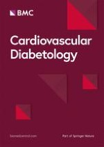
The formation of coronary collateral vessels (arteriogenesis) is a compensatory mechanism secondary to repetitive or chronic epicardial arterial blockage by arterial vasospasm or thrombosis. The process of arteriogenesis (collateral vessel formation) begins with small (20 micron diameter vessels) preexisting, quiescent, minimally – functioning arterioles and remodels them into larger (1–2 millimeter diameter vessels) more functional arterioles capable of carrying more oxygen enriched blood to ischemic tissue (a mini auto-coronary artery bypass). The shear stress associated with a pressure gradient as a result of epicardial coronary vasospasm or thrombosis is responsible for inducing the arteriolar remodeling process. In contrast, angiogenesis begins with capillaries and ends with more capillaries (figure 6,7). Important in this remodeling process are the shared cytokines, chemokines, growth factors, the inflammatory monocyte, and the extracellular matrix metalloproteases (MMPs) (table 4) [10].
Figure 6
Thespiritof vascularization. This figure compares and contrasts the involved mechanisms of angiogenesis and arteriogenesis. S = substrates, P = promotors, I = inducers, R = results, I = the common role of inflammation, and T = time. This acronym helps to understand why angiogenesis is induced and arteriogenesis is impaired.
Figure 7
Impairment of remodeling collateralization due to PAI-1. PAI-1 elevations impair fibrinolysis and contribute to a prothrombotic state in MS, PD, and T2DM. Additionally PAI-1 elevations impair Arteriogenesis. Remodeling collateralization is impaired in MS, PD, and T2DM due to elevations in PAI-1. Conversion of plasminogen to plasmin by tPA – uPA is negatively effected by elevations in PAI-1. This defect in the generation of Plasmin results in decreased conversion of latent MMPs to active MMPs and impair the remodeling necessary for the required remodeling of the preexisting arterioles to larger arterioles necessary for a more functional blood flow around obstructed epicardial coronary arteries and obstructed peripheral vascular systems in MS, PD, and T2DM.
(0) neutral Neutral effect: In Plaque. Ischemia "Shoulder" Macrophage and Hif-1 → VEGF override. Net effect neutral Plaque Angiogenesis remains induced: See Promoter and Inducer above.
(-) IMPAIRS Impairs Remodeling Collateralization due to negative effects on MMP activation and impaired ECM clearance for the remodeling collateralization mechanism.
ROS Increased in Diabetes
(+) increases SYNERGISTIC (to the above) SPIRIT of vascularization
(-) IMPAIRS ? Neutral to Negative in Collateral Vessels: See Promoter and Inducer above. Shear Stress → eNOS upregulation and eNO may override the negative effect of redox stress.
In the eNOS knockout model: Collateral formation was not impaired and the decreased flow could be restored with exogenous NO. This area of study needs further evaluation.
Oxygen Content
HYPOXIA
NORMOXIA
Recently Waltenberger J et al. were able to delineate the importance of the monocyte. They were able to demonstrate that monocyte migration was impaired in diabetes and associated with impaired collateral formation [37, 38]. Panutsopulos D et al. were also able to demonstrate an impairment of monocyte migration with an associated increase in basic fibroblast growth factor (bFGF) and vascular endothelial growth factor (VEGF) mRNA [39]. While the monocyte is an important cell associated with arteriogenesis and its impairment in migration will have a detrimental effect on remodeling collateralization there may also be a defect associated with MS, PD, and T2DM affecting the remodeling of the extracellular matrix (ECM).
MS, PD, and T2DM are associated with increased PAI-1 [8‐10]. PAI-1 inhibits both tissue and urokinase plasminogen activator resulting in impaired synthesis of plasmin. Plasmin is known to be very important in activating latent MMPs to active MMPs [40]. Therefore, PAI-1 excess may additionally serve to impair monocyte migration in addition to inhibiting the ECM remodeling collateralization (intima – media) of preexisting arterioles to the newly remodeled collateral arterioles (figure 7) [10].
There is a diabetes vascularization paradox in that angiogenesis is induced and arteriogenesis is impaired.
The diabetes vascularization paradox: accelerated intraplaque angiogenesis and impaired arteriogenesis
Angiogenesis and arteriogenesis are two distinct forms of postnatal vasculogenesis. Angiogenesis is induced by ischemia through the promoter Hif-1 TACGTGCT promoter and VEGF activity, whereas arteriogenesis is induced by the shear stress promotor by the shear stress responsive element (SSRE): GAGACC (figure 6) [41].
The atherosclerotic plaque angiogenic adventitial Vv within atheroscleropathy is excessive and associated with MS, PD, and overt T2DM. The Vv are induced by ischemia as the intima-media undergoes positive outward remodeling. Additionally, the Vv angiogenesis is further induced by the inflammatory process within the shoulder region of the plaque, which is associated with the known angiogenic factors: Tissue ACE, tissue factor, cytokines and growth factors (TNF alpha, VEGF, FGF). MS, PD, and T2DM are associated with multiple metabolic toxicities, which are responsible for reactive oxygen species (ROS) production (table 3). This elevated tension of redox stress contributes to the ischemia within the plaque and induces even more angiogenesis within the plaque.
Table 3
Multiple metabolic toxicities in ms and t2dm: the a-flight acronym
Initiator
Metabolic Defect
Metabolic mediator
Functional mediator
Consequence ROS
A
AMYLIN
(Co-secreted – Co-packaged within the insulin secretory granule) by the islet Beta cell.
Insulin's "Fraternal Twin" Elevated in MS, PD, and Early T2DM)
REDOX STRESS Activated Platelets PAI-1 elevation Fibrinogen elevated. Decreased NO
ROS Athero-emboli ROS
In contrast arteriogenesis is induced and promoted by shear stress in normoxic conditions (table 4). Redox stress and ROS are elevated in the preexisting (un-remodeled) arteriolar endothelial milieu in MS, PD, and T2DM and is associated with endothelial dysfunction with decreased eNOS activity and decreased generation of eNO. This could result in an impaired initial vasodilatation and permeability ordinarily induced by increased shear stress in the arteriogenic process. Terjung RL et al. and Matsunaga T et al. have reported that arteriogenesis is eNOS and eNO dependent [42, 43]. Thus, the impairment in eNOS and eNO (due to redox stress and ROS) [8] could impair the initiation and progression of arteriogenesis and result in a negative effect on remodeling collateralization.
Given that collateral formation is inhibited by NOS inhibition, dysfunction or knockout, one can hypothesize that eNOS dysfunction and – or decreased eNO may decrease arteriogenesis through the following mechanism: a decrease in arteriolar vasodilation would impair the normal arteriolar vasodilation due to eNOS and eNO and decrease the pressure gradient between normal and ischemic tissue. This would impair the normal increased flow and the shear stress responsive element in the collateral vessel undergoing remodeling collateralization, as well as, decreasing endothelial cell permeability [44]. Future work is needed to establish if reversing endothelial dysfunction associated with MS, PD, and T2DM could restore improved arteriogenesis [10]. Thus it appears that a blunted endothelial NO production as noted in MS, PD, and overt T2DM could temper vascular remodeling and arteriogenesis [45].
Excess in redox stress or ROS and PAI-1 elevation could have an overall negative effect on arteriogenesis in MS, PD, and overt T2DM (table 4).
Another possibility is the existence of early advanced glycation end products (AGE) and that these AGE would contribute not only to the impaired remodeling collateralization phase but also contribute to additional collagen cross linking within the extracellular matrix. If glucotoxicity were to be better controlled this might improve the remodeling phase of arteriogenesis in T2DM by improving redox stress, endothelial dysfunction, monocyte migration, and PAI-1 levels.
Endotheliopathy
MS, PD, and T2DM are associated with a diffuse endotheliopathy. This endothelial dysfunction can be related to the three vulnerable arms of the endothelial nitric oxide synthase (eNOS) reaction.
Oxidative – redox stress is elevated in MS, PD, and T2DM and results in the production of reactive oxygen species (ROS). The multiple metabolic toxicities of the A-FLIGHT acronym result in an abundance of ROS. These ROS interfere with the eNOS reaction resulting in a decrease in endothelial nitric oxide (eNO). The process of uncoupling of the eNOS reaction allows this reaction to proceed with the endothelium becoming a net producer of superoxide [O2•] instead of the quintessential, protective, anti-inflammatory, antioxidant eNO. When L-arginine uncouples from the eNOS enzyme via an intact necessary cofactor tetrahydrobiopterin (BH4) the following reaction ensues:
The BH4 requisite cofactor itself is quite sensitive to oxidative – redox stress and can be oxidized to BH2 and BH3, which will not run the eNOS reaction and allow uncoupling. This allows the endothelium to become a net producer of superoxide instead of eNO. As can be seen it is very important to have an adequate substrate, a proper functioning eNOS enzyme, and the necessary cofactor BH4. Folic acid is not only a methyl donor and aids in the control of elevated homocysteine but also an electron and hydrogen donor, which brings BH2 and BH3 back to the requisite completely reduced BH4. These findings would lead one to not only control the multiple metabolic toxicities (A-FLIGHT table 3) but also provide folic acid in adequate amounts to allow BH4 to run the eNOS reaction fully re-coupled to once again become a net producer of eNO [8, 9].
Conclusion
MS, PD, and T2DM are associated with endothelial dysfunction and carry an elevated risk for both micro and macrovascular disease that are often present at the time of diagnosis of overt T2DM. Postnatal vascularization in a response to injury mechanism to the endothelium and the arterial vessel wall as well as the capillary bed result in most of the complications associated with these disorders.
This review has focused on the arterial vessel wall in order to better understand the development and acceleration of macrovascular disease termed: atheroscleropathy and the capillary bed in microvascular disease. The diabetic vascularization paradox has been presented in order to better understand the mechanisms involved in the finding of angiogenesis being induced and arteriogenesis impaired.
The important role of plaque angiogenesis and subsequent plaque destabilization has been presented as this mechanism plays such an important role in plaque rupture and acute coronary syndromes. The epicardial coronary vessels and the peripheral arterial vessels are involved with ischemic manifestations as a result of atheroscleropathy and impaired arteriogenesis – collateral vascular formation.
When you stand back and examine the overall vascular health in MS, PD, and T2DM it becomes obvious that treatment to prevent, slow, or halt these vascular abnormalities requires a global risk reduction approach in treatment in order to treat the multiple metabolic toxic abnormalities associated with this complex disease (figure 2) (table 3). We currently have multiple treatment regimens available to use as each metabolic abnormality is addressed clinically. The RAAS acronym is provided as a tool to assist the clinician in the treatment of these complicated multiple metabolic derangements (table 5). Each therapeutic component of the RAAS acronym will have a positive effect on the excessive angiogenesis and the impaired arteriogenesis associated with MS and T2DM atheroscleropathy [46‐48].
Table 5
THE RAAS ACRONYM: GLOBAL RISK REDUCTION
R
Reductase inhibitors (HMG-CoA). Decreasing modified LDL-cholesterol, i.e. oxidized, acetylated LDL-cholesterol. Decreasing triglycerides and increasing HDL-cholesterol Improving endothelial cell dysfunction. Restoring the abnormal Lipoprotein fractions. Thus, decreasing the redox and oxidative stress to the arterial vessel wall and myocardium.
Redox stress reduction.
A
AngII inhibition or blockade:
ACE inhibitors – Angiotensin II receptor blockers: Both inhibiting the effect of angiotensin-II locally as well as systemically. Affecting hemodynamic stress through their antihypertensive effect as well as the deleterious effects of angiotensin II on cells at the local level – injurious stimuli-decreasing the stimulus for O2•production. Decreasing the A-FLIGHT toxicities. Plus the direct-indirect antioxidant effect within the arterial vessel wall and capillary. Antioxidant effects.
Aspirin antiplatelet, anti-inflammatory effect.
Adrenergic (non-selective blockade) in addition to its blockade of Prorenin → Renin
Amlodipine with its calcium channel blocking antihypertensive effect, in addition to its direct antioxidant effects.
Redox stress reduction.
A
Aggressive control of diabetes to HbA1c of less than 7. (This usually requires combination therapy with the use of: Insulin secretagogues, insulin sensitizers (thiazolidinediones), biguanides, alpha-glucosidase inhibitors, and ultimately exogenous insulin.).
Decreasing modified LDL cholesterol, i.e. glycated – glycoxidated LDL cholesterol. Improving endothelial cell dysfunction. Also decreasing glucotoxicity and the oxidative – redox stress to the intima and pancreatic islet.
Aggressive control of blood pressure, which usually requires combination therapy, including thiazide diuretics to attain JNC 7 guidelines.
Aggressive control of Hcy with folic acid and its associated pleiotropic positive effect on re-coupling the eNOS reaction by restoring the activity of the BH4 cofactor to run the eNOS reaction and once again produce eNO, as well as, its direct antioxidant effects: BH4 and eNOS stabilization
Redox stress reduction.
S
Statins. Improving plaque stability (pleiotropic effects) independent of cholesterol lowering. Improving endothelial cell dysfunction. Plus, the direct – indirect antioxidant anti-inflammatory effects [45] within the islet and the arterial vessel wall promoting stabilization of the unstable, vulnerable islet and the arterial vessel wall. Style: Lifestyle modification: lose weight, exercise, and change eating habits. S top S moking
Nicotine Adenine Di nucleotide Phosphate reduced oxidase.
AGE
Advanced Glycation Endproducts.
AFE
Advanced Fructosylation Endproducts.
RAGE
Receptor for Advanced Glycosylation Endproducts.
ALE
Advanced Lipoxidation Endproducts.
eNOS
Endothelial Nitric Oxide Synthase.
NO
Nitric Oxide.
BH4
Tetra Hydro Biopterin.
FFA
Free Fatty Acids.
LC acyl -CoA's
Long chain Acyl Co enzyme CoA.
VLDL
Very low density lipoprotein.
LDL
Low density lipoprotein.
HDL
High density lipoprotein.
MS
Metabolic Syndrome.
PD
Prediabetes.
T2DM
Type 2 Diabetes Mellitus.
PAI-1
Plasminogen Activator Inhibitor-1.
H2O
Water.
Glut-4
Glucose Transporter-4.
PI3 Kinase
Phosotidyl inositol 3 Kinase.
Akt
Protein kinase B.
MAP Kinase
Mitogen Activated Protein Kinase.
MAP Kinase Shunt
MAP Kinase Shunt: The shunting away from the positive Glut 4 PI3 Kinase Akt pathway to the deleterious MAP Kinase pathway promoting remodeling due to an alteration in the NO redox sensitive PI3 Kinase /Akt pathway.
___
IL-6 IL-8
Interleukin-6 Interleukin-8.
TNF alpha
Tumor Necrosis Factor alpha.
MPO
Myeloperoxidase: Generation of Superoxide (O2•) via hypochlorous acid HClO-
NF kappa B
Nuclear Factor kappa B.
ICAM
Inter Cellular Adhesion Molecule.
VCAM,
Vascular Cellular Adhesion Molecule.
MCP-1
Monocyte Chemoattractant Protein-1
NADH
Nicotinamide Adenine Dinucleotide reduced
NAD+
Nicotinamide Adenine Dinucleotide oxidized
DAG
Diacylglycerol.
GPx
Glutathione Peroxidase.
DDAH
Dimethylarginine dimethylaminohydrolase.
ADMA
Asymmetrical dimethyl arginine.
O2•- ONOO•
Superoxide – Peroxynitrite.
Authors contributions
M. R. Hayden and S.C. Tyagi contributed equally in the inception, writing, and editing of this manuscript.
Acknowledgements
A part of this study was supported by NIH grants HL-71010 and HL-74185.
The authors wish to acknowledge the late A. Cliff Barger and Jeffrey M. Isner for their large body of work they have contributed to the importance of angiogenesis in the field of cardiovascular disease. Also, to acknowledge the large body of work Wolfgang Schaper and colleagues have contributed to the importance of arteriogenesis in the field of cardiovascular disease.
Ein signifikanter Anteil der Fälle von plötzlichem Herztod ist genetisch bedingt. Um ihre Verwandten vor diesem Schicksal zu bewahren, sollten jüngere Personen, die plötzlich unerwartet versterben, ausnahmslos einer Autopsie unterzogen werden.
Kommt es zu einer nichttraumatischen Hirnblutung, spielt es keine große Rolle, ob die Betroffenen zuvor direkt wirksame orale Antikoagulanzien oder Marcumar bekommen haben: Die Prognose ist ähnlich schlecht.
Nicht nur ein vergrößerter, sondern auch ein kleiner linker Ventrikel ist bei Vorhofflimmern mit einer erhöhten Komplikationsrate assoziiert. Der Zusammenhang besteht nach Daten aus China unabhängig von anderen Risikofaktoren.
Bei adipösen Patienten mit Herzinsuffizienz des HFpEF-Phänotyps ist Semaglutid von symptomatischem Nutzen. Resultiert dieser Benefit allein aus der Gewichtsreduktion oder auch aus spezifischen Effekten auf die Herzinsuffizienz-Pathogenese? Eine neue Analyse gibt Aufschluss.
Update Innere Medizin
Bestellen Sie unseren Fach-Newsletter und bleiben Sie gut informiert.